CLARITY in Alzheimer’s Research: Merging Tissue Transparency with Next-Gen Neurotechnologies
IF 2.3
4区 医学
Q2 BIOCHEMICAL RESEARCH METHODS
引用次数: 0
Abstract
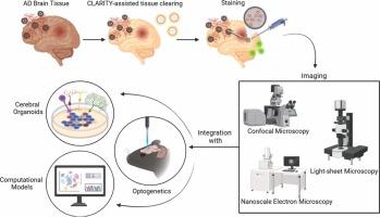
CLARITY is a technique that makes tissues optically transparent, enabling the clear visualization of complex cellular and subcellular structures with relative ease. Traditionally, this technique has been used to visualize the pathologies of certain diseases. In the case of Alzheimer’s disease (AD), the CLARITY technique of clearing lipids from tissues has enabled precise visualization of amyloid-beta (Aβ) and tau pathologies with a temporal analysis of the extent of protein aggregation associated with disease progression. This information has been invaluable for determining the progression of AD. Moreover, the structural characteristics of Aβ plaques and neurofibrillary tangles provide details about aggregation patterns. Here, we highlight critical insights into the potential applications of the CLARITY technique in conjunction with other modalities. We elucidated microscopy techniques, namely confocal microscopy and light-sheet microscopy, which are often used to detect fluorescently labeled compounds within cells, aiding in the qualitative and quantitative estimation of target proteins. Additionally, we discuss the application of super-resolution expansion microscopy (SREM) to increase the resolution of tissue images without compromising molecular specificity. We also expounded on the application of CLARITY-based imaging in improving optogenetics and organoid development. Furthermore, we highlight the potential of CLARITY in assisting the formulation and validation of machine learning algorithms and conducting hypothesis testing.
阿尔茨海默病研究的清晰度:将组织透明度与新一代神经技术相结合。
CLARITY是一种使组织具有光学透明性的技术,能够相对容易地清晰地显示复杂的细胞和亚细胞结构。传统上,这项技术已被用于可视化某些疾病的病理。在阿尔茨海默病(AD)的病例中,清除组织脂质的CLARITY技术能够精确地可视化淀粉样蛋白- β (a β)和tau病理,并对与疾病进展相关的蛋白质聚集程度进行时间分析。这一信息对于确定阿尔茨海默病的进展是非常宝贵的。此外,Aβ斑块和神经原纤维缠结的结构特征提供了聚集模式的细节。在这里,我们强调了CLARITY技术与其他模式结合的潜在应用的关键见解。我们阐明了显微镜技术,即共聚焦显微镜和光片显微镜,它们通常用于检测细胞内荧光标记的化合物,有助于靶蛋白的定性和定量估计。此外,我们讨论了超分辨率扩展显微镜(SREM)的应用,以提高组织图像的分辨率,而不影响分子特异性。并阐述了基于清晰度的成像技术在改善光遗传学和类器官发育方面的应用。此外,我们强调了CLARITY在协助机器学习算法的制定和验证以及进行假设检验方面的潜力。
本文章由计算机程序翻译,如有差异,请以英文原文为准。
求助全文
约1分钟内获得全文
求助全文
来源期刊

Journal of Neuroscience Methods
医学-神经科学
CiteScore
7.10
自引率
3.30%
发文量
226
审稿时长
52 days
期刊介绍:
The Journal of Neuroscience Methods publishes papers that describe new methods that are specifically for neuroscience research conducted in invertebrates, vertebrates or in man. Major methodological improvements or important refinements of established neuroscience methods are also considered for publication. The Journal''s Scope includes all aspects of contemporary neuroscience research, including anatomical, behavioural, biochemical, cellular, computational, molecular, invasive and non-invasive imaging, optogenetic, and physiological research investigations.
 求助内容:
求助内容: 应助结果提醒方式:


