{"title":"The Impact of Helicobacter pylori Infection on Retinal Nerve Fiber Layer and Macula.","authors":"Hui Miao, TianHao Qu, Yanming Huang, Sujie Fan, Yiquan Yang, Aiguo Lv, Jianhua Hu, Li Guo, Qian Jia, Zhenduo Li, Shujun Zhang, Yuhong Li, Wei Shi, Yongjie Chen, Siyue Chen, Hua Yan","doi":"10.1167/iovs.66.11.35","DOIUrl":null,"url":null,"abstract":"<p><strong>Purpose: </strong>This study aims to elucidate the effects of Helicobacter pylori infection on the retinal nerve fiber layer (RNFL) and macula.</p><p><strong>Methods: </strong>A cross-sectional study was undertaken at the Health Examination Center of the First Hospital of Handan from 2021 to 2022. Participants who underwent both the 14C-urea breath test and ocular examinations were selected. The RNFL and macula were assessed using a swept-source optical coherence tomography device (Topcon OCT, Tokyo, Japan). Data are presented as mean ± SD and were analyzed using the Mann-Whitney U-test and t-test, with a significance level set at P < 0.05.</p><p><strong>Results: </strong>RNFL analysis of 1693 H. pylori-positive and 1693 negative left eyes revealed significantly thinner inferior temporal RNFL in the H. pylori-positive group (149.79 ± 29.55 µm vs. 152.76 ± 29.15 µm, P = 0.026). No other regional RNFL differences were found (P > 0.05). In the macula study of 2117 H. pylori-positive and 2117 negative participants, macular degeneration occurred in 74 positive (avgerage age 54.66 ± 10.08 years) and 67 negative individuals (avgerage age 60.22 ± 10.50 years). The incidence rates were not significantly different (P > 0.05), but the positive group with lesions was significantly younger (P = 0.002).</p><p><strong>Conclusions: </strong>H. pylori infection may be associated with localized defects in the RNFL, which could serve as early indicators of glaucoma, and it may also be potentially linked to accelerated progression of macular degeneration.</p>","PeriodicalId":14620,"journal":{"name":"Investigative ophthalmology & visual science","volume":"66 11","pages":"35"},"PeriodicalIF":4.7000,"publicationDate":"2025-08-01","publicationTypes":"Journal Article","fieldsOfStudy":null,"isOpenAccess":false,"openAccessPdf":"https://www.ncbi.nlm.nih.gov/pmc/articles/PMC12366866/pdf/","citationCount":"0","resultStr":null,"platform":"Semanticscholar","paperid":null,"PeriodicalName":"Investigative ophthalmology & visual science","FirstCategoryId":"3","ListUrlMain":"https://doi.org/10.1167/iovs.66.11.35","RegionNum":2,"RegionCategory":"医学","ArticlePicture":[],"TitleCN":null,"AbstractTextCN":null,"PMCID":null,"EPubDate":"","PubModel":"","JCR":"Q1","JCRName":"OPHTHALMOLOGY","Score":null,"Total":0}
引用次数: 0
Abstract
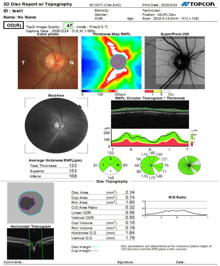
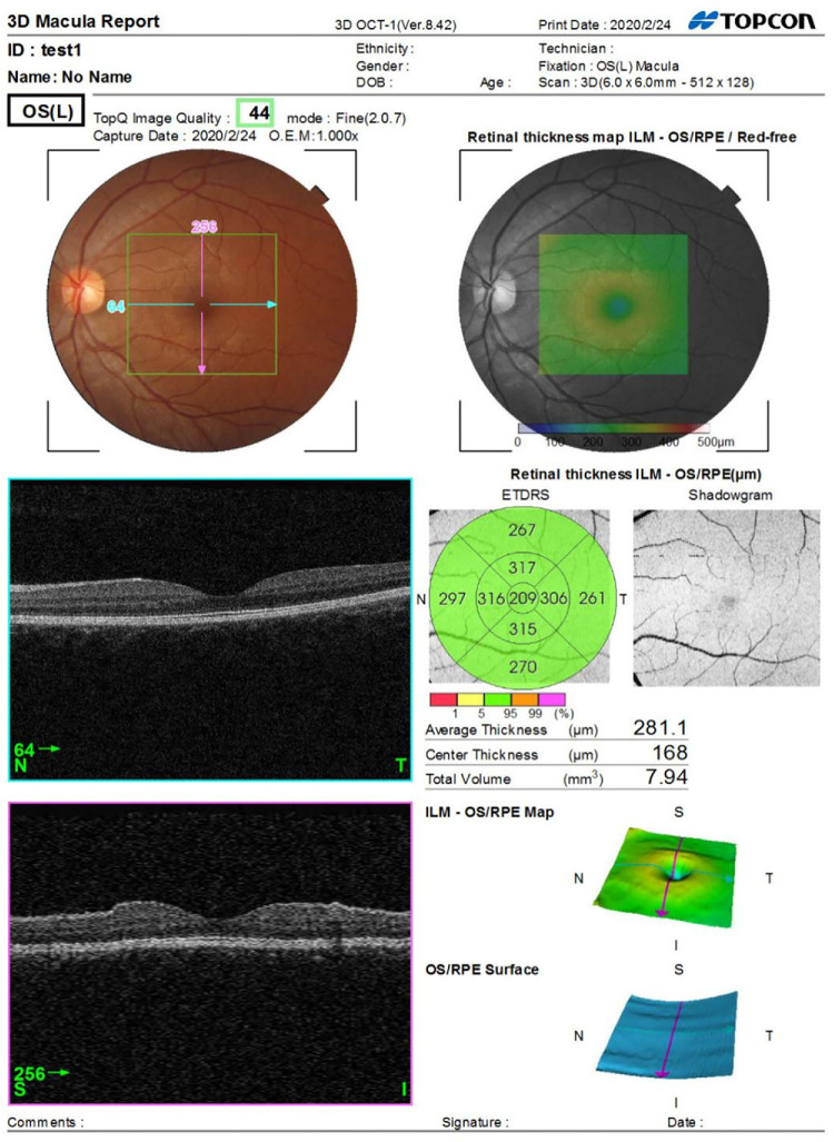
Purpose: This study aims to elucidate the effects of Helicobacter pylori infection on the retinal nerve fiber layer (RNFL) and macula.
Methods: A cross-sectional study was undertaken at the Health Examination Center of the First Hospital of Handan from 2021 to 2022. Participants who underwent both the 14C-urea breath test and ocular examinations were selected. The RNFL and macula were assessed using a swept-source optical coherence tomography device (Topcon OCT, Tokyo, Japan). Data are presented as mean ± SD and were analyzed using the Mann-Whitney U-test and t-test, with a significance level set at P < 0.05.
Results: RNFL analysis of 1693 H. pylori-positive and 1693 negative left eyes revealed significantly thinner inferior temporal RNFL in the H. pylori-positive group (149.79 ± 29.55 µm vs. 152.76 ± 29.15 µm, P = 0.026). No other regional RNFL differences were found (P > 0.05). In the macula study of 2117 H. pylori-positive and 2117 negative participants, macular degeneration occurred in 74 positive (avgerage age 54.66 ± 10.08 years) and 67 negative individuals (avgerage age 60.22 ± 10.50 years). The incidence rates were not significantly different (P > 0.05), but the positive group with lesions was significantly younger (P = 0.002).
Conclusions: H. pylori infection may be associated with localized defects in the RNFL, which could serve as early indicators of glaucoma, and it may also be potentially linked to accelerated progression of macular degeneration.
期刊介绍:
Investigative Ophthalmology & Visual Science (IOVS), published as ready online, is a peer-reviewed academic journal of the Association for Research in Vision and Ophthalmology (ARVO). IOVS features original research, mostly pertaining to clinical and laboratory ophthalmology and vision research in general.

 求助内容:
求助内容: 应助结果提醒方式:


