Felix Schlachetzki, Ina Feistenauer, Michael Ertl, Mustafa Kilic, Fabian Aden, David Pollinger, Horst Helbig, Christina Wendl, Karin Pfister, Lars Krenkel, Maria Andreea Gamulescu, Ralf Andreas Linker, Sibylle Wilfling
{"title":"Retinal ischemia due to different stages of atherosclerosis - insights from a retrospective study on central retinal artery occlusion.","authors":"Felix Schlachetzki, Ina Feistenauer, Michael Ertl, Mustafa Kilic, Fabian Aden, David Pollinger, Horst Helbig, Christina Wendl, Karin Pfister, Lars Krenkel, Maria Andreea Gamulescu, Ralf Andreas Linker, Sibylle Wilfling","doi":"10.1186/s42466-025-00413-z","DOIUrl":null,"url":null,"abstract":"<p><strong>Background: </strong>Ischemic stroke (IS) and retinal ischemia (IR) share similar vascular risk factors, but differ in their risk for subsequent or recurrent stroke and therapeutic options. This study characterizes the cardiovascular risk profiles and magnitude of atherosclerosis of the carotid artery of patients with central retinal artery occlusion (CRAO) in relation to the presence of the retrobulbar \"spot sign\" on orbital color-coded sonography (OCCS).</p><p><strong>Methods: </strong>We performed a retrospective analysis on the detailed cardiovascular risk factors and neuroimaging data in patients with IR presenting between 2009 and 2023. Based on OCCS findings, CRAO were further divided into hyperechoic (\"spot sign positive\", ssCRAO) or hypoechoic CRAO (heCRAO). Statistical analyses were performed with Mann-Whitney-U and χ [2] testing. P-values were considered significant if < 0.05.</p><p><strong>Results: </strong>Overall, 112 patients were identified (heCRAO: n = 32; ssCRAO: n = 80). ssCRAO patients were significantly older (median 74 years vs. 66.5 years, Mann-Whitney-U: p-value < 0.001). Overall, 15/103 (14.6%) patients had concurrent acute ischemic stroke- 9 in the ipsilateral internal carotid territory, 2 in other territories and 4 disseminated. Further significant differences were found regarding the echogenicity of atherosclerosis (AS) in the two subgroups with (mainly) echorich AS being more common in the ssCRAO group (p-value < 0.001, n = 108) and the distribution of high-grade vs. low-grade stenoses of the ipsi- and contralateral carotid artery (p-value < 0.05, n = 99). 20 out of 112 patients had atrial fibrillation (aFib) with 17 of these being on ongoing oral anticoagulation.</p><p><strong>Conclusion: </strong>According to this study, atherosclerosis may be one of the most important risk factors for IR while a specific embolic source could not be demonstrated (i.e. acute plaque rupture). By contrast, current oral anticoagulation for aFib in CRAO patients was high, thus only an incidental finding and may be an incidental finding due to its prevalence in the elderly. Furthermore, we were able to distinguish two subgroups of IR that differ in risk factors and most likely also in etiology, therapy and prognosis. The study underlines the importance of OCCS to detect \"spot signs\" in IR with indications for both, acute thrombolysis and secondary prevention.</p>","PeriodicalId":94156,"journal":{"name":"Neurological research and practice","volume":"7 1","pages":"50"},"PeriodicalIF":3.2000,"publicationDate":"2025-07-22","publicationTypes":"Journal Article","fieldsOfStudy":null,"isOpenAccess":false,"openAccessPdf":"https://www.ncbi.nlm.nih.gov/pmc/articles/PMC12285047/pdf/","citationCount":"0","resultStr":null,"platform":"Semanticscholar","paperid":null,"PeriodicalName":"Neurological research and practice","FirstCategoryId":"1085","ListUrlMain":"https://doi.org/10.1186/s42466-025-00413-z","RegionNum":0,"RegionCategory":null,"ArticlePicture":[],"TitleCN":null,"AbstractTextCN":null,"PMCID":null,"EPubDate":"","PubModel":"","JCR":"Q2","JCRName":"Medicine","Score":null,"Total":0}
引用次数: 0
Abstract
Background: Ischemic stroke (IS) and retinal ischemia (IR) share similar vascular risk factors, but differ in their risk for subsequent or recurrent stroke and therapeutic options. This study characterizes the cardiovascular risk profiles and magnitude of atherosclerosis of the carotid artery of patients with central retinal artery occlusion (CRAO) in relation to the presence of the retrobulbar "spot sign" on orbital color-coded sonography (OCCS).
Methods: We performed a retrospective analysis on the detailed cardiovascular risk factors and neuroimaging data in patients with IR presenting between 2009 and 2023. Based on OCCS findings, CRAO were further divided into hyperechoic ("spot sign positive", ssCRAO) or hypoechoic CRAO (heCRAO). Statistical analyses were performed with Mann-Whitney-U and χ [2] testing. P-values were considered significant if < 0.05.
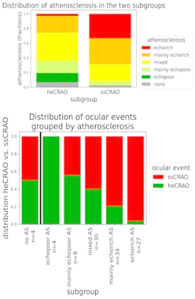
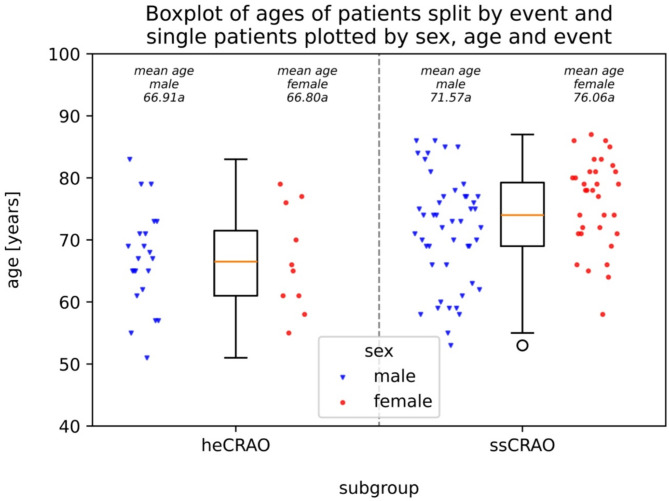
Results: Overall, 112 patients were identified (heCRAO: n = 32; ssCRAO: n = 80). ssCRAO patients were significantly older (median 74 years vs. 66.5 years, Mann-Whitney-U: p-value < 0.001). Overall, 15/103 (14.6%) patients had concurrent acute ischemic stroke- 9 in the ipsilateral internal carotid territory, 2 in other territories and 4 disseminated. Further significant differences were found regarding the echogenicity of atherosclerosis (AS) in the two subgroups with (mainly) echorich AS being more common in the ssCRAO group (p-value < 0.001, n = 108) and the distribution of high-grade vs. low-grade stenoses of the ipsi- and contralateral carotid artery (p-value < 0.05, n = 99). 20 out of 112 patients had atrial fibrillation (aFib) with 17 of these being on ongoing oral anticoagulation.
Conclusion: According to this study, atherosclerosis may be one of the most important risk factors for IR while a specific embolic source could not be demonstrated (i.e. acute plaque rupture). By contrast, current oral anticoagulation for aFib in CRAO patients was high, thus only an incidental finding and may be an incidental finding due to its prevalence in the elderly. Furthermore, we were able to distinguish two subgroups of IR that differ in risk factors and most likely also in etiology, therapy and prognosis. The study underlines the importance of OCCS to detect "spot signs" in IR with indications for both, acute thrombolysis and secondary prevention.
背景:缺血性脑卒中(IS)和视网膜缺血(IR)具有相似的血管危险因素,但其后续或复发性脑卒中的风险和治疗选择不同。本研究描述了视网膜中央动脉闭塞(CRAO)患者的心血管风险特征和颈动脉粥样硬化程度与眼眶彩色超声(OCCS)上球后“斑点征”的存在之间的关系。方法:我们对2009年至2023年间IR患者的详细心血管危险因素和神经影像学资料进行回顾性分析。根据OCCS结果,CRAO进一步分为高回声(“斑点标志阳性”,ssCRAO)或低回声CRAO (heCRAO)。采用Mann-Whitney-U和χ[2]检验进行统计分析。如果结果如下,则认为p值具有显著性:总体而言,鉴定出112例患者(heCRAO: n = 32;ssCRAO: n = 80)。结论:根据这项研究,动脉粥样硬化可能是IR最重要的危险因素之一,而具体的栓塞来源无法证实(即急性斑块破裂)。相比之下,目前在CRAO患者中口服抗凝治疗aFib的比例很高,因此只是一个偶然发现,可能是偶然发现,因为它在老年人中流行。此外,我们能够区分两个不同的IR亚组,它们在危险因素上不同,很可能在病因、治疗和预后上也不同。该研究强调了OCCS在急性溶栓和二级预防适应症中检测IR“斑点征象”的重要性。

 求助内容:
求助内容: 应助结果提醒方式:


