Mohammad Yousefi, Nicola Maffulli, Marjan Bahraminasab, Samaneh Arab, Akram Alizadeh, Ali Ghanbari, Athar Talebi, Muhammad Mehdi Jafari Sorkhdehi
{"title":"Critical-size bone defect repair with three types of nano-hydroxyapatite scaffolds: An <i>in vivo</i> study.","authors":"Mohammad Yousefi, Nicola Maffulli, Marjan Bahraminasab, Samaneh Arab, Akram Alizadeh, Ali Ghanbari, Athar Talebi, Muhammad Mehdi Jafari Sorkhdehi","doi":"10.34172/bi.30247","DOIUrl":null,"url":null,"abstract":"<p><p></p><p><strong>Introduction: </strong>Hydroxyapatite (HA), the main mineral component of bone, can be synthesized and utilized in the bone lesion treatments because of its high bioactivity and osteoconductive property. HA extraction from fish bones has received special attention given its low cost and easier extraction protocol compared to other sources. The present study compared the biocompatibility and bone repair of commercial nano hydroxyapatite (nHA) powder with that extracted from carp and human bones <i>in vitro</i> and <i>in vivo</i>.</p><p><strong>Methods: </strong>First, nHA powders were prepared, and their physical and structural properties were studied using XRD, FTIR, FE-SEM and EDS analyses. Next, the powders were used to make porous scaffolds for which the physicochemical, structural, mechanical and biological properties were evaluated. The <i>in vitro</i> biocompatibility and osteogenic differentiation were tested on MC3T3-E1 cells, respectively, by MTT assay in three time periods and Alizarin red staining. Furthermore, the scaffolds were implanted after creation of critical-size lesions in the skulls of female rats, and the histological investigation was conducted by H&E staining at two time points.</p><p><strong>Results: </strong>The morphological and phase analyses showed the successful fabrication of porous nHA scaffolds with 60.68%, 61.38, and 63.27% for carp, human and commercial nHA scaffolds, respectively. The scaffolds showed different biodegradability behavior where the human nHA scaffolds degrade more rapidly. The results of mechanical tests indicated that the scaffolds made up of human extracted nHA powder had the lowest strength and stiffness (3.13 and 37.37 KPa, respectively). The strength and stiffness of the scaffolds fabricated by carp extracted and commercial nHA were 17.14 and 19.01 Kpa, and 251.79 and 140.49 Kpa, respectively. The MTT test results showed that the greatest cell viability rate was in the carp nHA scaffolds after 10 days (146.08%). Moreover, the AR staining indicated the formation of mineralized nodules caused by the scaffolds in all groups. However, the mineralization seemed to be superior in human, and carp extracted groups. Furthermore, <i>in vivo</i> in all three groups bone repair occurred at the critical-size lesion sites, while scaffolds biodegradation was also observed. The scaffolds made up of carp and human nHA exhibited the highest rate of ossification and maturation of bone tissue among different scaffolds after 8 weeks. The rate of tissue response to these scaffolds was higher than the scaffolds made of commercial nHA after 4 and 8 weeks, postoperatively.</p><p><strong>Conclusion: </strong>The carp extracted nHA scaffolds perform comparable to human extracted nHA, and may be used for clinical applications.</p>","PeriodicalId":48614,"journal":{"name":"Bioimpacts","volume":"15 ","pages":"30247"},"PeriodicalIF":2.2000,"publicationDate":"2025-03-01","publicationTypes":"Journal Article","fieldsOfStudy":null,"isOpenAccess":false,"openAccessPdf":"https://www.ncbi.nlm.nih.gov/pmc/articles/PMC12204782/pdf/","citationCount":"0","resultStr":null,"platform":"Semanticscholar","paperid":null,"PeriodicalName":"Bioimpacts","FirstCategoryId":"5","ListUrlMain":"https://doi.org/10.34172/bi.30247","RegionNum":4,"RegionCategory":"工程技术","ArticlePicture":[],"TitleCN":null,"AbstractTextCN":null,"PMCID":null,"EPubDate":"2025/1/1 0:00:00","PubModel":"eCollection","JCR":"Q3","JCRName":"PHARMACOLOGY & PHARMACY","Score":null,"Total":0}
引用次数: 0
Abstract
Introduction: Hydroxyapatite (HA), the main mineral component of bone, can be synthesized and utilized in the bone lesion treatments because of its high bioactivity and osteoconductive property. HA extraction from fish bones has received special attention given its low cost and easier extraction protocol compared to other sources. The present study compared the biocompatibility and bone repair of commercial nano hydroxyapatite (nHA) powder with that extracted from carp and human bones in vitro and in vivo.
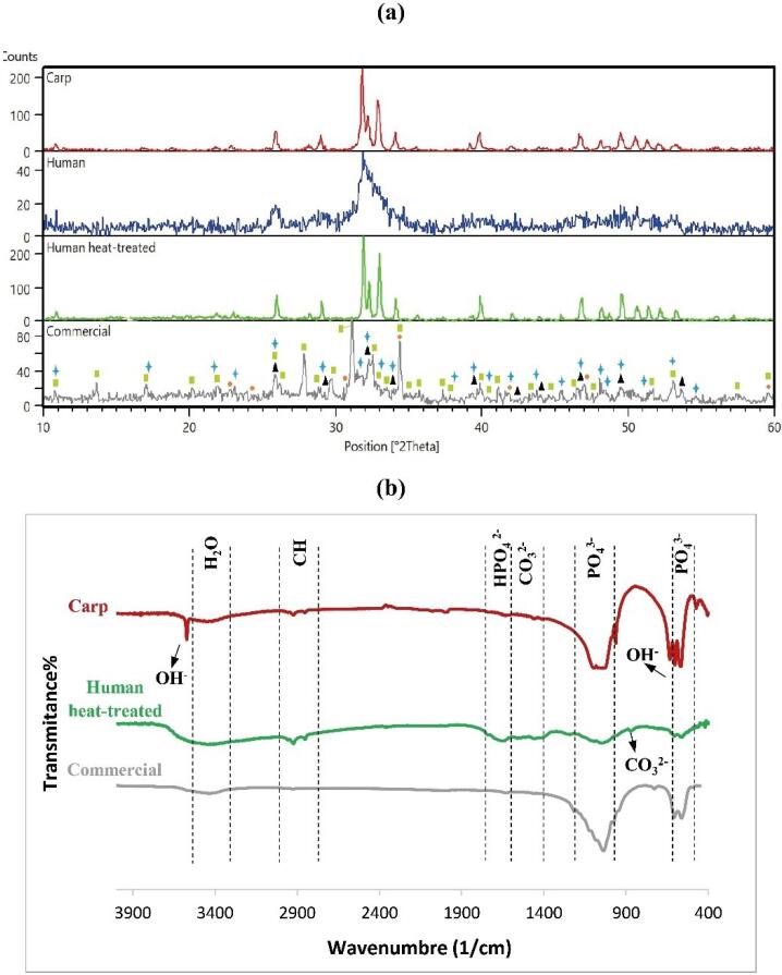
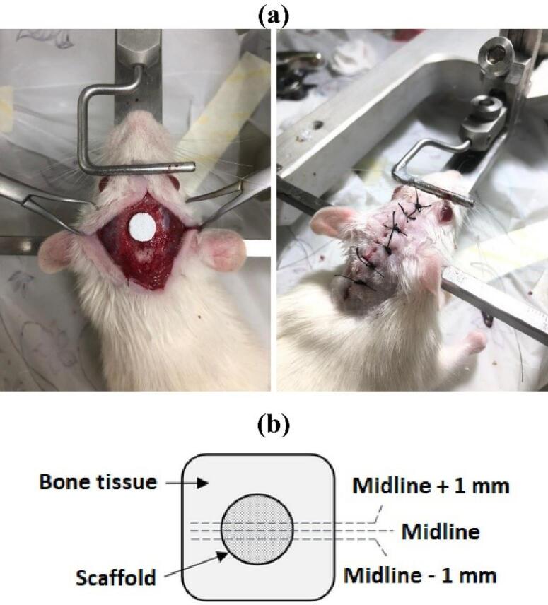
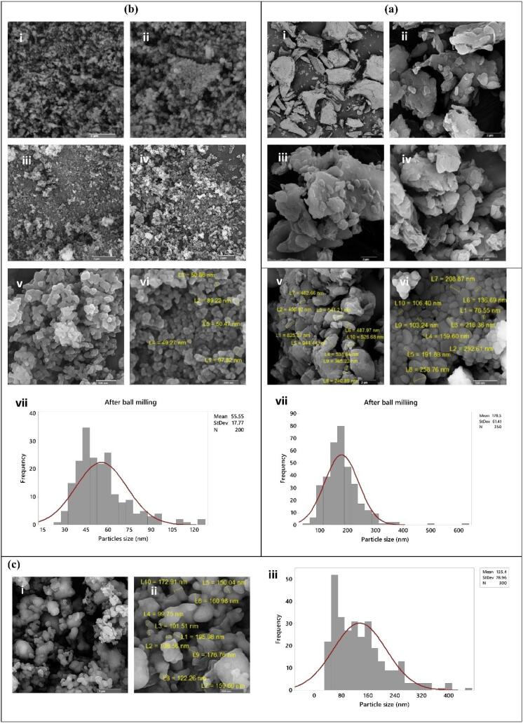
Methods: First, nHA powders were prepared, and their physical and structural properties were studied using XRD, FTIR, FE-SEM and EDS analyses. Next, the powders were used to make porous scaffolds for which the physicochemical, structural, mechanical and biological properties were evaluated. The in vitro biocompatibility and osteogenic differentiation were tested on MC3T3-E1 cells, respectively, by MTT assay in three time periods and Alizarin red staining. Furthermore, the scaffolds were implanted after creation of critical-size lesions in the skulls of female rats, and the histological investigation was conducted by H&E staining at two time points.
Results: The morphological and phase analyses showed the successful fabrication of porous nHA scaffolds with 60.68%, 61.38, and 63.27% for carp, human and commercial nHA scaffolds, respectively. The scaffolds showed different biodegradability behavior where the human nHA scaffolds degrade more rapidly. The results of mechanical tests indicated that the scaffolds made up of human extracted nHA powder had the lowest strength and stiffness (3.13 and 37.37 KPa, respectively). The strength and stiffness of the scaffolds fabricated by carp extracted and commercial nHA were 17.14 and 19.01 Kpa, and 251.79 and 140.49 Kpa, respectively. The MTT test results showed that the greatest cell viability rate was in the carp nHA scaffolds after 10 days (146.08%). Moreover, the AR staining indicated the formation of mineralized nodules caused by the scaffolds in all groups. However, the mineralization seemed to be superior in human, and carp extracted groups. Furthermore, in vivo in all three groups bone repair occurred at the critical-size lesion sites, while scaffolds biodegradation was also observed. The scaffolds made up of carp and human nHA exhibited the highest rate of ossification and maturation of bone tissue among different scaffolds after 8 weeks. The rate of tissue response to these scaffolds was higher than the scaffolds made of commercial nHA after 4 and 8 weeks, postoperatively.
Conclusion: The carp extracted nHA scaffolds perform comparable to human extracted nHA, and may be used for clinical applications.
BioimpactsPharmacology, Toxicology and Pharmaceutics-Pharmaceutical Science
CiteScore
4.80
自引率
7.70%
发文量
36
审稿时长
5 weeks
期刊介绍:
BioImpacts (BI) is a peer-reviewed multidisciplinary international journal, covering original research articles, reviews, commentaries, hypotheses, methodologies, and visions/reflections dealing with all aspects of biological and biomedical researches at molecular, cellular, functional and translational dimensions.
 求助内容:
求助内容: 应助结果提醒方式:


