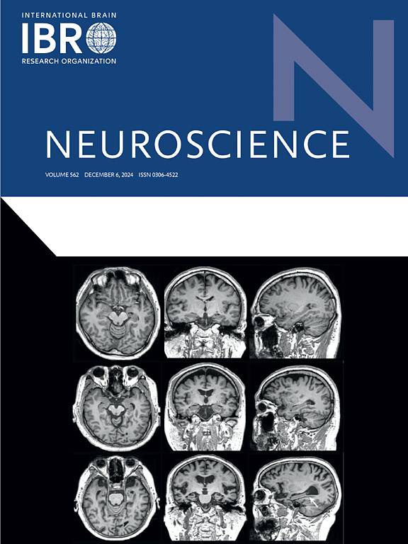
Audio-visual crossmodal connectivity in perceiving distorted speech: A comparison between patients with cleft palate and typical listeners
IF 2.8
3区 医学
Q2 NEUROSCIENCES
引用次数: 0
Abstract
This study investigates audio-visual crossmodal connectivity during the perception of distorted speech using task-based functional magnetic resonance imaging (fMRI). A randomized block design was employed, involving 20 patients with cleft palate and 20 typical listeners. Participants underwent perceptual tasks involving both cleft-related glottal stop and typical speech while fMRI scans were conducted. Regional interactions between the auditory and visual cortices were analyzed using dynamic causal modeling (DCM). Individual-level effective connectivity analysis revealed that, during the perception of glottal stop, patients with cleft palate exhibited significantly reduced effective connectivity from the left superior temporal gyrus to the left inferior occipital gyrus compared to typical listeners (p = 0.035). However, no significant difference was observed in the connectivity weights from the right superior temporal gyrus to the right inferior occipital gyrus. These findings suggest a potential deficit in audio-visual integration in patients with cleft palate, which may adversely affect speech perception. This insight advances our understanding of the neural mechanisms underlying speech disorders in cleft palate, particularly the contribution of crossmodal connectivity to speech processing.
腭裂患者与典型听者感知扭曲言语的视听跨模态连通性比较
本研究利用任务型功能磁共振成像(fMRI)研究了语音失真感知过程中的视听跨模态连接。采用随机区组设计,选取20例腭裂患者和20例典型听者。在进行功能磁共振成像扫描的同时,参与者接受了包括与唇裂相关的声门停顿和典型语音的感知任务。利用动态因果模型(DCM)分析了听觉和视觉皮层之间的区域相互作用。个体水平的有效连通性分析显示,在声门停止的感知过程中,腭裂患者的左颞上回到左枕下回的有效连通性明显低于典型听者(p = 0.035)。而右颞上回与右枕下回的连通性权重无显著差异。这些发现表明腭裂患者可能存在潜在的视听整合缺陷,这可能对言语感知产生不利影响。这一发现促进了我们对腭裂言语障碍的神经机制的理解,特别是跨模态连接对言语处理的贡献。
本文章由计算机程序翻译,如有差异,请以英文原文为准。
求助全文
约1分钟内获得全文
求助全文
来源期刊

Neuroscience
医学-神经科学
CiteScore
6.20
自引率
0.00%
发文量
394
审稿时长
52 days
期刊介绍:
Neuroscience publishes papers describing the results of original research on any aspect of the scientific study of the nervous system. Any paper, however short, will be considered for publication provided that it reports significant, new and carefully confirmed findings with full experimental details.
 求助内容:
求助内容: 应助结果提醒方式:


