Randa Obid, Austin R. Green, Sion W. Jasmine, Rachel P. Kowal, Brooj Abro, Laura M. Warmke, Magdalena B. Czader, Ahmed K. Alomari, Carina A. Dehner
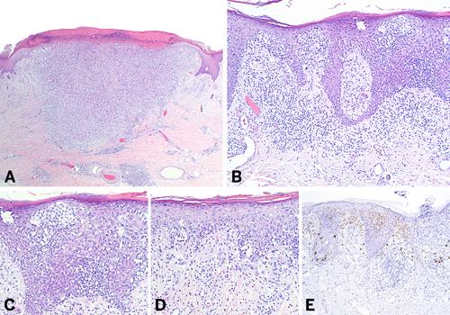
{"title":"An Unusual Case of Cutaneous Langerhans Cell Sarcoma Lacking S100 Expression: A Case Report and Review of the Literature","authors":"Randa Obid, Austin R. Green, Sion W. Jasmine, Rachel P. Kowal, Brooj Abro, Laura M. Warmke, Magdalena B. Czader, Ahmed K. Alomari, Carina A. Dehner","doi":"10.1111/cup.14833","DOIUrl":null,"url":null,"abstract":"<p>Langerhans cell sarcoma (LCS) is a rare neoplastic proliferation of Langerhans cell with aggressive clinical behavior and involves multiple organ systems, including the skin. LCS is characterized by marked cytologic atypia, frequent mitoses including atypical ones, and expression of CD1a, S100, and langerin (CD207). CD1a and Langerin-positive but S100− negative LCS is extremely rare in clinical practice. We present a case of a 71-year-old female with a history of melanoma and atypical fibroxanthoma who presented with an erythematous plaque on her left knee. Histopathologic examination revealed a dermal infiltrate comprised of large pleomorphic cells with irregular nuclear contours, prominent longitudinal grooves, and vesicular chromatin, and a high mitotic rate. Notably, there were epidermotropism and a distinctive immunohistochemical profile: S100−, CD1a+, Langerin+, and focal CD68+. Next-generation sequencing identified copy number loss of <i>CDKN2A</i>, <i>CDKN2B</i>, and <i>FOXA1</i>, mutations in <i>TP53</i>, <i>POT1</i>, <i>SH2B3</i>, and <i>SMARCA4</i>, and a high tumor mutational burden. Herein, we discuss the clinical and pathologic features of 38 cases of LCS with cutaneous involvement reported in the literature, including recent advances in understanding molecular characteristics of this disease. This exceptional case may contribute to our understanding of the etiology of this rare neoplasm.</p>","PeriodicalId":15407,"journal":{"name":"Journal of Cutaneous Pathology","volume":"52 9","pages":"554-567"},"PeriodicalIF":1.1000,"publicationDate":"2025-06-24","publicationTypes":"Journal Article","fieldsOfStudy":null,"isOpenAccess":false,"openAccessPdf":"https://onlinelibrary.wiley.com/doi/epdf/10.1111/cup.14833","citationCount":"0","resultStr":null,"platform":"Semanticscholar","paperid":null,"PeriodicalName":"Journal of Cutaneous Pathology","FirstCategoryId":"3","ListUrlMain":"https://onlinelibrary.wiley.com/doi/10.1111/cup.14833","RegionNum":4,"RegionCategory":"医学","ArticlePicture":[],"TitleCN":null,"AbstractTextCN":null,"PMCID":null,"EPubDate":"","PubModel":"","JCR":"Q3","JCRName":"DERMATOLOGY","Score":null,"Total":0}
引用次数: 0
Abstract
Langerhans cell sarcoma (LCS) is a rare neoplastic proliferation of Langerhans cell with aggressive clinical behavior and involves multiple organ systems, including the skin. LCS is characterized by marked cytologic atypia, frequent mitoses including atypical ones, and expression of CD1a, S100, and langerin (CD207). CD1a and Langerin-positive but S100− negative LCS is extremely rare in clinical practice. We present a case of a 71-year-old female with a history of melanoma and atypical fibroxanthoma who presented with an erythematous plaque on her left knee. Histopathologic examination revealed a dermal infiltrate comprised of large pleomorphic cells with irregular nuclear contours, prominent longitudinal grooves, and vesicular chromatin, and a high mitotic rate. Notably, there were epidermotropism and a distinctive immunohistochemical profile: S100−, CD1a+, Langerin+, and focal CD68+. Next-generation sequencing identified copy number loss of CDKN2A, CDKN2B, and FOXA1, mutations in TP53, POT1, SH2B3, and SMARCA4, and a high tumor mutational burden. Herein, we discuss the clinical and pathologic features of 38 cases of LCS with cutaneous involvement reported in the literature, including recent advances in understanding molecular characteristics of this disease. This exceptional case may contribute to our understanding of the etiology of this rare neoplasm.
期刊介绍:
Journal of Cutaneous Pathology publishes manuscripts broadly relevant to diseases of the skin and mucosae, with the aims of advancing scientific knowledge regarding dermatopathology and enhancing the communication between clinical practitioners and research scientists. Original scientific manuscripts on diagnostic and experimental cutaneous pathology are especially desirable. Timely, pertinent review articles also will be given high priority. Manuscripts based on light, fluorescence, and electron microscopy, histochemistry, immunology, molecular biology, and genetics, as well as allied sciences, are all welcome, provided their principal focus is on cutaneous pathology. Publication time will be kept as short as possible, ensuring that articles will be quickly available to all interested in this speciality.

 求助内容:
求助内容: 应助结果提醒方式:


