Stephanie Quinn, Vasileios T Papastavrou, Clare Warriner, Jill M O'Brien, Michael E Grinton, Andrew C Browning
{"title":"Early retinal electrophysiology changes in quinine overdose.","authors":"Stephanie Quinn, Vasileios T Papastavrou, Clare Warriner, Jill M O'Brien, Michael E Grinton, Andrew C Browning","doi":"10.1007/s10633-025-10034-1","DOIUrl":null,"url":null,"abstract":"<p><strong>Purpose: </strong>To report the early and subsequent electrophysiological findings of 2 patients following quinine overdose.</p><p><strong>Methods: </strong>Serial assessments including: Medical history, visual acuity (VA), fundus autofluorescence, spectral-domain macular optical coherence tomography (OCT) and full-field electroretinogram (ffERG) were performed on 2 patients, between 2 and 47 days after quinine overdose.</p><p><strong>Results: </strong>Both patients experienced a similar clinical course. After almost total vision loss within the first 24 h, VA dramatically improved by day 3. Early OCT changes demonstrated central macula hyperautofluorescence, which coincided with a hyperreflectivity of the macular inner retina on OCT. The initial ffERG findings demonstrated changes consistent with marked inner retinal dysfunction of the cone system, affecting both the cone ON- and OFF-bipolar cell pathways. In contrast, rod bipolar cell function was unaffected in the early phase of toxicity. Between days 10 and 17, the retinal arterioles showed narrowing which coincided with attenuation of ffERG parameters of rod system inner retinal function between days 10-40.</p><p><strong>Conclusions: </strong>These cases suggest the early stages of quinine toxicity affect function of the presynaptic cone bipolar cell junction. This is then followed by retinal arteriolar attenuation and the well described electronegative scotopic ffERG.</p>","PeriodicalId":11207,"journal":{"name":"Documenta Ophthalmologica","volume":" ","pages":"179-186"},"PeriodicalIF":2.9000,"publicationDate":"2025-10-01","publicationTypes":"Journal Article","fieldsOfStudy":null,"isOpenAccess":false,"openAccessPdf":"https://www.ncbi.nlm.nih.gov/pmc/articles/PMC12436528/pdf/","citationCount":"0","resultStr":null,"platform":"Semanticscholar","paperid":null,"PeriodicalName":"Documenta Ophthalmologica","FirstCategoryId":"3","ListUrlMain":"https://doi.org/10.1007/s10633-025-10034-1","RegionNum":4,"RegionCategory":"医学","ArticlePicture":[],"TitleCN":null,"AbstractTextCN":null,"PMCID":null,"EPubDate":"2025/6/17 0:00:00","PubModel":"Epub","JCR":"Q2","JCRName":"OPHTHALMOLOGY","Score":null,"Total":0}
引用次数: 0
Abstract
Purpose: To report the early and subsequent electrophysiological findings of 2 patients following quinine overdose.
Methods: Serial assessments including: Medical history, visual acuity (VA), fundus autofluorescence, spectral-domain macular optical coherence tomography (OCT) and full-field electroretinogram (ffERG) were performed on 2 patients, between 2 and 47 days after quinine overdose.
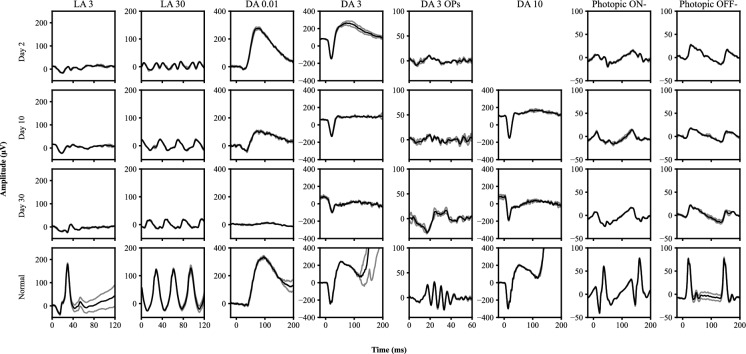
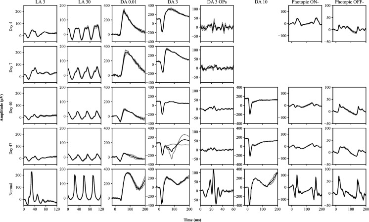
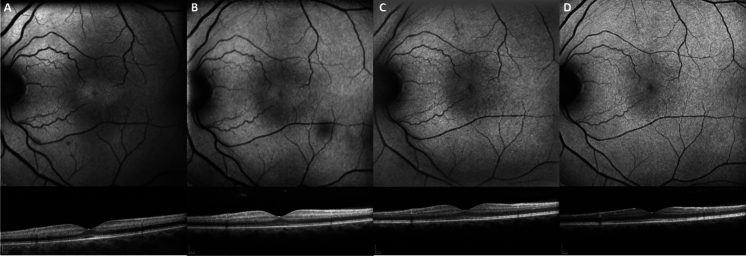
Results: Both patients experienced a similar clinical course. After almost total vision loss within the first 24 h, VA dramatically improved by day 3. Early OCT changes demonstrated central macula hyperautofluorescence, which coincided with a hyperreflectivity of the macular inner retina on OCT. The initial ffERG findings demonstrated changes consistent with marked inner retinal dysfunction of the cone system, affecting both the cone ON- and OFF-bipolar cell pathways. In contrast, rod bipolar cell function was unaffected in the early phase of toxicity. Between days 10 and 17, the retinal arterioles showed narrowing which coincided with attenuation of ffERG parameters of rod system inner retinal function between days 10-40.
Conclusions: These cases suggest the early stages of quinine toxicity affect function of the presynaptic cone bipolar cell junction. This is then followed by retinal arteriolar attenuation and the well described electronegative scotopic ffERG.
期刊介绍:
Documenta Ophthalmologica is an official publication of the International Society for Clinical Electrophysiology of Vision. The purpose of the journal is to promote the understanding and application of clinical electrophysiology of vision. Documenta Ophthalmologica will publish reviews, research articles, technical notes, brief reports and case studies which inform the readers about basic and clinical sciences related to visual electrodiagnosis and means to improve diagnosis and clinical management of patients using visual electrophysiology. Studies may involve animals or humans. In either case appropriate care must be taken to follow the Declaration of Helsinki for human subject or appropriate humane standards of animal care (e.g., the ARVO standards on Animal Care and Use).

 求助内容:
求助内容: 应助结果提醒方式:


