Imaging-based maxillomandibular changes after head and neck radiotherapy: A systematic review and meta-analysis
IF 2
3区 医学
Q2 DENTISTRY, ORAL SURGERY & MEDICINE
Journal of Stomatology Oral and Maxillofacial Surgery
Pub Date : 2025-10-01
DOI:10.1016/j.jormas.2025.102432
引用次数: 0
Abstract
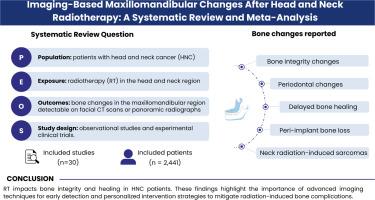
This systematic review aims to evaluate the maxillomandibular bone changes post-radiotherapy in head and neck cancer (HNC) patients using qualitative and quantitative imaging analyses. The review was conducted following PRISMA guidelines, using the PEOS framework: patients with HNC (population), radiotherapy in the head and neck region (exposure), bone changes in the jaws detectable by radiographs (outcomes), including observational studies and experimental clinical trials (study design). A comprehensive search was performed in PubMed, Embase, Cochrane, and Web of Science, along with gray literature. Demographic data, tumor characteristics, radiotherapy modalities, and imaging findings from computed tomography scans or panoramic radiographs were extracted, with the risk of bias assessed using JBI’s critical appraisal tools, and studies at high risk of bias were excluded from the meta-analysis. A total of 30 studies involving 2441 patients met the inclusion criteria. Bone integrity alterations were the most frequently reported outcomes, including osteoradionecrosis (ORN), bone sclerosis, cortical thinning, trabecular density reduction, and osteolytic and osteosclerotic lesions. ORN was observed in 7 % of cases (95 % CI: 4–10 %). Periodontal alterations, such as periodontal ligament widening, were reported in five studies. Impaired bone healing included delayed socket ossification and nonunion after surgery. Panoramic radiography was the most used imaging modality, while quantitative analyses, such as fractal dimension reductions and radiomics, revealed subtle changes indicative of bone deterioration. Radiotherapy significantly impacts bone integrity and healing in HNC patients. These findings highlight the importance of advanced imaging techniques for early detection and personalized intervention strategies to mitigate radiation-induced bone complications.
头颈部放疗后基于成像的上颌骨变化:系统回顾和荟萃分析。
本系统综述旨在通过定性和定量影像学分析评估头颈癌(HNC)患者放射治疗后上颌骨的变化。该综述遵循PRISMA指南,使用PEOS框架:HNC患者(人群)、头颈部放疗(暴露)、x线片检测到的颌骨骨变化(结果),包括观察性研究和实验性临床试验(研究设计)。我们在PubMed、Embase、Cochrane和Web of Science以及灰色文献中进行了全面的搜索。提取人口统计学数据、肿瘤特征、放疗方式以及计算机断层扫描或全景x线片的成像结果,使用JBI的关键评估工具评估偏倚风险,并将高偏倚风险的研究排除在meta分析之外。共有30项涉及2441例患者的研究符合纳入标准。骨完整性改变是最常见的报道结果,包括骨放射性坏死(ORN)、骨硬化、皮质变薄、小梁密度降低、溶骨和骨硬化病变。7%的病例出现ORN (95% CI: 4-10%)。牙周改变,如牙周韧带扩大,在五项研究中被报道。骨愈合受损包括手术后窝骨化延迟和骨不连。全景x线摄影是最常用的成像方式,而定量分析,如分形维数降低和放射组学,显示了指示骨恶化的细微变化。放疗显著影响HNC患者的骨完整性和愈合。这些发现强调了先进的成像技术对早期发现和个性化干预策略的重要性,以减轻辐射引起的骨并发症。
本文章由计算机程序翻译,如有差异,请以英文原文为准。
求助全文
约1分钟内获得全文
求助全文
来源期刊

Journal of Stomatology Oral and Maxillofacial Surgery
Surgery, Dentistry, Oral Surgery and Medicine, Otorhinolaryngology and Facial Plastic Surgery
CiteScore
2.30
自引率
9.10%
发文量
0
审稿时长
23 days
 求助内容:
求助内容: 应助结果提醒方式:


