Gwladys Revêchon, Anna Witasp, Nikenza Viceconte, Hafdis T. Helgadottir, Piotr Machtel, Fabiana Stefani, Daniel Whisenant, Agustin Sola-Carvajal, Dagmara McGuinness, Nadia O. Abutaleb, Gonzalo Artiach, Emelie Wallén Arzt, Inga Soveri, Anne Babler, Susanne Ziegler, Rafael Kramann, Magnus Bäck, Anders Thorell, George A. Truskey, Lars Wennberg, Paul G. Shiels, Annika Wernerson, Peter Stenvinkel, Maria Eriksson
{"title":"Recurrent somatic mutation and progerin expression in early vascular aging of chronic kidney disease","authors":"Gwladys Revêchon, Anna Witasp, Nikenza Viceconte, Hafdis T. Helgadottir, Piotr Machtel, Fabiana Stefani, Daniel Whisenant, Agustin Sola-Carvajal, Dagmara McGuinness, Nadia O. Abutaleb, Gonzalo Artiach, Emelie Wallén Arzt, Inga Soveri, Anne Babler, Susanne Ziegler, Rafael Kramann, Magnus Bäck, Anders Thorell, George A. Truskey, Lars Wennberg, Paul G. Shiels, Annika Wernerson, Peter Stenvinkel, Maria Eriksson","doi":"10.1038/s43587-025-00882-6","DOIUrl":null,"url":null,"abstract":"Early vascular aging plays a central role in chronic kidney disease (CKD), but its molecular causes remain unclear. Somatic mutations accumulate in various cells with age, yet their functional contribution to aging tissues is not well understood. Here we found progerin, the protein responsible for the premature aging disease Hutchinson–Gilford progeria syndrome, steadily recurring in vascular smooth muscle cells of patients with CKD. Notably, the most common progeria-causing mutation, LMNA c.1824C>T, was identified as a somatic mutation in CKD arteries. Clusters of proliferative progerin-expressing cells in CKD arteries and in vivo lineage-tracing in mice revealed clonal expansion capacity of mutant cells. Mosaic progerin expression contributed to genomic damage, endoplasmic reticulum stress and senescence in CKD arteries and resulted in vascular aging phenotypes in vivo. These findings suggest that certain somatic mutations may be clonally expanded in the arterial wall, contributing to the disease-related functional decline of the tissue. Progerin, a truncated version of lamin A, causes Hutchinson–Gilford progeria syndrome characterized by premature aging and cardiovascular complications. Here the authors show that the most common progeria-causing mutation, LMNA c.1824C>T, is a somatic mutation in arteries from patients with chronic kidney disease, and that clonal propagation of the mutation in the vascular wall results in vascular aging phenotypes in mice.","PeriodicalId":94150,"journal":{"name":"Nature aging","volume":"5 6","pages":"1046-1062"},"PeriodicalIF":19.4000,"publicationDate":"2025-06-10","publicationTypes":"Journal Article","fieldsOfStudy":null,"isOpenAccess":false,"openAccessPdf":"https://www.ncbi.nlm.nih.gov/pmc/articles/PMC12176630/pdf/","citationCount":"0","resultStr":null,"platform":"Semanticscholar","paperid":null,"PeriodicalName":"Nature aging","FirstCategoryId":"1085","ListUrlMain":"https://www.nature.com/articles/s43587-025-00882-6","RegionNum":0,"RegionCategory":null,"ArticlePicture":[],"TitleCN":null,"AbstractTextCN":null,"PMCID":null,"EPubDate":"","PubModel":"","JCR":"Q1","JCRName":"CELL BIOLOGY","Score":null,"Total":0}
引用次数: 0
Abstract
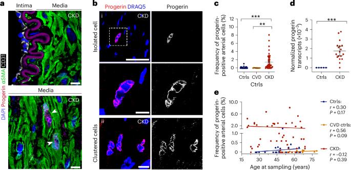
Early vascular aging plays a central role in chronic kidney disease (CKD), but its molecular causes remain unclear. Somatic mutations accumulate in various cells with age, yet their functional contribution to aging tissues is not well understood. Here we found progerin, the protein responsible for the premature aging disease Hutchinson–Gilford progeria syndrome, steadily recurring in vascular smooth muscle cells of patients with CKD. Notably, the most common progeria-causing mutation, LMNA c.1824C>T, was identified as a somatic mutation in CKD arteries. Clusters of proliferative progerin-expressing cells in CKD arteries and in vivo lineage-tracing in mice revealed clonal expansion capacity of mutant cells. Mosaic progerin expression contributed to genomic damage, endoplasmic reticulum stress and senescence in CKD arteries and resulted in vascular aging phenotypes in vivo. These findings suggest that certain somatic mutations may be clonally expanded in the arterial wall, contributing to the disease-related functional decline of the tissue. Progerin, a truncated version of lamin A, causes Hutchinson–Gilford progeria syndrome characterized by premature aging and cardiovascular complications. Here the authors show that the most common progeria-causing mutation, LMNA c.1824C>T, is a somatic mutation in arteries from patients with chronic kidney disease, and that clonal propagation of the mutation in the vascular wall results in vascular aging phenotypes in mice.

 求助内容:
求助内容: 应助结果提醒方式:


