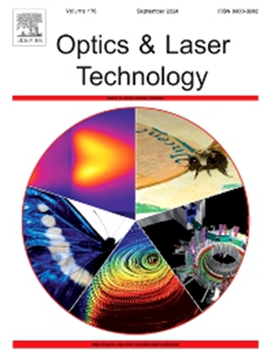
Improved shape from focus network for extended depth of field and rapid three-dimensional reconstruction biological microscopy
IF 4.6
2区 物理与天体物理
Q1 OPTICS
引用次数: 0
Abstract
Three-dimensional (3D) reconstruction microscopy has played an important role in advancing the elucidation of the roles and structures of biological cells, but the current mainstream optical microimaging techniques make it difficult to capture the 3D structures of dynamic organisms. Therefore, this paper proposes a fast and versatile end-to-end improved shape-from-focus (ISFF) network and enlarged selective kernel (ESK) module, which are applied to obtain microscopes with a large depth of field and high-precision 3D reconstruction capability. To characterize the feasibility of ISFF, our algorithm achieves a higher quality of the fused image compared to six prevalent deep learning image fusion algorithms. We apply our microscopy to 3D imaging of live biological samples such as bee tentacles, C.elegans, and zebrafish without fluorescent labels or anesthesia, and our experimental results show that high-resolution 3D observation of biodynamic processes can be achieved in 1.86 s. Quantitative analysis of the interface between the standard gauge blocks shows that the microscopy’s depth of field extends to 1200 μm under a 10 × objective and the relative errors of the reconstruction for the two gauge blocks are 0.61 % and 0.54 %, respectively. Our network eliminates the need to train exclusively on model organisms such as C. elegans and zebrafish, while still achieving good 3D reconstruction results. It not only expands the application range and system robustness of biomicroscopy but also provides new perspectives and tools for living model organisms at the millimeter scale.
改进的形状从焦点网络扩展景深和快速三维重建生物显微镜
三维重建显微技术在推动生物细胞功能和结构的阐明方面发挥了重要作用,但目前主流的光学微成像技术难以捕捉动态生物体的三维结构。为此,本文提出了一种快速通用的端到端改进聚焦形状(ISFF)网络和放大选择核(ESK)模块,用于获得具有大景深和高精度三维重建能力的显微镜。为了表征ISFF的可行性,与六种流行的深度学习图像融合算法相比,我们的算法实现了更高的融合图像质量。我们将显微镜应用于活体生物样品的三维成像,如蜜蜂触手、秀丽隐杆线虫和斑马鱼,没有荧光标记或麻醉,我们的实验结果表明,生物动力学过程的高分辨率三维观察可以在1.86 s内实现。对标准规块之间的界面进行了定量分析,结果表明,在10倍物镜下,显微镜的景深可达1200 μm,两个规块的重建相对误差分别为0.61%和0.54%。我们的网络消除了只对秀丽隐杆线虫和斑马鱼等模式生物进行训练的需要,同时仍然可以获得良好的3D重建结果。它不仅扩大了生物显微镜的应用范围和系统鲁棒性,而且为毫米尺度的活体模式生物提供了新的视角和工具。
本文章由计算机程序翻译,如有差异,请以英文原文为准。
求助全文
约1分钟内获得全文
求助全文
来源期刊
CiteScore
8.50
自引率
10.00%
发文量
1060
审稿时长
3.4 months
期刊介绍:
Optics & Laser Technology aims to provide a vehicle for the publication of a broad range of high quality research and review papers in those fields of scientific and engineering research appertaining to the development and application of the technology of optics and lasers. Papers describing original work in these areas are submitted to rigorous refereeing prior to acceptance for publication.
The scope of Optics & Laser Technology encompasses, but is not restricted to, the following areas:
•development in all types of lasers
•developments in optoelectronic devices and photonics
•developments in new photonics and optical concepts
•developments in conventional optics, optical instruments and components
•techniques of optical metrology, including interferometry and optical fibre sensors
•LIDAR and other non-contact optical measurement techniques, including optical methods in heat and fluid flow
•applications of lasers to materials processing, optical NDT display (including holography) and optical communication
•research and development in the field of laser safety including studies of hazards resulting from the applications of lasers (laser safety, hazards of laser fume)
•developments in optical computing and optical information processing
•developments in new optical materials
•developments in new optical characterization methods and techniques
•developments in quantum optics
•developments in light assisted micro and nanofabrication methods and techniques
•developments in nanophotonics and biophotonics
•developments in imaging processing and systems

 求助内容:
求助内容: 应助结果提醒方式:


