{"title":"Dermoscopic Features of Eccrine Poromas in Diverse Skin Phototypes: A Retrospective Study of 26 Cases.","authors":"Bengu Nisa Akay, Handan Merve Erol Mart, Aylin Okcu Heper","doi":"10.5826/dpc.1502a5071","DOIUrl":null,"url":null,"abstract":"<p><strong>Introduction: </strong>Eccrine poroma (EP) is a benign adnexal tumor. Establishing a definitive diagnosis based on clinical and dermoscopic findings can be challenging.</p><p><strong>Objectives: </strong>The aim of this study was to perform a comprehensive analysis of the dermoscopic features of pigmented, hypopigmented, and nonpigmented variants of EP and to compare these dermoscopic features in patients with dark and light Fitzpatrick skin phototypes.</p><p><strong>Methods: </strong>A total of 26 cases of histopathologically confirmed EP were included. Each case was categorized as pigmented, hypopigmented, or nonpigmented based on the melanin content within the lesion. Patients were classified according to their Fitzpatrick skin phototypes. Dermoscopic images were subjected to revised pattern analysis, and the results were compared with the existing literature.</p><p><strong>Results: </strong>Regarding Fitzpatrick skin phototype, four (15.4%), 11 (42.3%), six (23.1%), and five (19.2%) patients had Fitzpatrick skin phototypes II, III, IV, and V, respectively. Of the cases, 17 (65.4%) were classified as nonpigmented, three (11.5%) as hypopigmented, and six (23.1%) as pigmented EP. All pigmented EP cases occurred in patients with dark skin and were located on non-acral sites. Polymorphic vascular pattern, branched vessels with rounded endings, linear-irregular vessels, interlacing white areas around vessels, and collarettes were more frequently observed in patients with light skin. In contrast, clod vessels, coiled vessels, white lines, ulceration, fiber sign, scales, and structureless areas were more common in patients with dark skin.</p><p><strong>Conclusions: </strong>This study underscores the significant dermoscopic diversity observed in EP, revealing distinct patterns based on pigmentation and Fitzpatrick skin phototypes.</p>","PeriodicalId":11168,"journal":{"name":"Dermatology practical & conceptual","volume":"15 2","pages":""},"PeriodicalIF":2.3000,"publicationDate":"2025-04-01","publicationTypes":"Journal Article","fieldsOfStudy":null,"isOpenAccess":false,"openAccessPdf":"https://www.ncbi.nlm.nih.gov/pmc/articles/PMC12090919/pdf/","citationCount":"0","resultStr":null,"platform":"Semanticscholar","paperid":null,"PeriodicalName":"Dermatology practical & conceptual","FirstCategoryId":"3","ListUrlMain":"https://doi.org/10.5826/dpc.1502a5071","RegionNum":4,"RegionCategory":"医学","ArticlePicture":[],"TitleCN":null,"AbstractTextCN":null,"PMCID":null,"EPubDate":"","PubModel":"","JCR":"Q2","JCRName":"DERMATOLOGY","Score":null,"Total":0}
引用次数: 0
Abstract
Introduction: Eccrine poroma (EP) is a benign adnexal tumor. Establishing a definitive diagnosis based on clinical and dermoscopic findings can be challenging.
Objectives: The aim of this study was to perform a comprehensive analysis of the dermoscopic features of pigmented, hypopigmented, and nonpigmented variants of EP and to compare these dermoscopic features in patients with dark and light Fitzpatrick skin phototypes.
Methods: A total of 26 cases of histopathologically confirmed EP were included. Each case was categorized as pigmented, hypopigmented, or nonpigmented based on the melanin content within the lesion. Patients were classified according to their Fitzpatrick skin phototypes. Dermoscopic images were subjected to revised pattern analysis, and the results were compared with the existing literature.
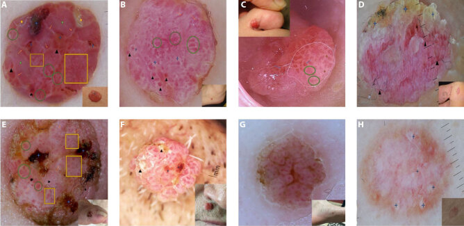
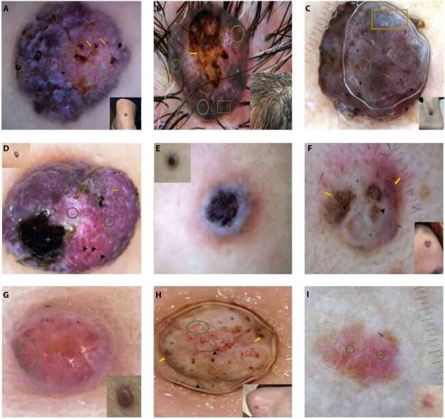
Results: Regarding Fitzpatrick skin phototype, four (15.4%), 11 (42.3%), six (23.1%), and five (19.2%) patients had Fitzpatrick skin phototypes II, III, IV, and V, respectively. Of the cases, 17 (65.4%) were classified as nonpigmented, three (11.5%) as hypopigmented, and six (23.1%) as pigmented EP. All pigmented EP cases occurred in patients with dark skin and were located on non-acral sites. Polymorphic vascular pattern, branched vessels with rounded endings, linear-irregular vessels, interlacing white areas around vessels, and collarettes were more frequently observed in patients with light skin. In contrast, clod vessels, coiled vessels, white lines, ulceration, fiber sign, scales, and structureless areas were more common in patients with dark skin.
Conclusions: This study underscores the significant dermoscopic diversity observed in EP, revealing distinct patterns based on pigmentation and Fitzpatrick skin phototypes.
 求助内容:
求助内容: 应助结果提醒方式:


