{"title":"Thalidomide augments maturation and T helper 1-inducing capacity of monocyte-derived dendritic cells in vitro.","authors":"Mohsen Abbaszadeh, Bahar Naseri, Javad Masoumi, Elham Baghbani, Behzad Baradaran, Mohammad Reza Sadeghi","doi":"10.34172/bi.30588","DOIUrl":null,"url":null,"abstract":"<p><p></p><p><strong>Introduction: </strong>Dendritic cells (DCs) possess specialized abilities to present antigens and stimulate T cells, making them essential in triggering adaptive immune responses. Thalidomide and its derivatives are classified as a group of medications that possess immunomodulatory properties. Numerous studies have demonstrated the contentious impact of these drugs on DCs. Therefore, the objective of the present study was to assess the influence of Thalidomide therapy on the maturation and stimulation of monocyte-derived DCs, and subsequently examine the consequences of these treated DCs on the immune responses of autologous T cells.</p><p><strong>Methods: </strong>The immature DCs derived from monocytes were subjected to exposure to Thalidomide and Lipopolysaccharides (LPS) on the fifth day of differentiation, followed by a 24-hour incubation period. On the sixth day, the phenotypic features of the DCs in both the control and treatment groups were assessed using flow cytometry. Subsequently, the gene expression in both the DCs and autologous T cells co-cultured with the DCs was evaluated using the real-time PCR method.</p><p><strong>Results: </strong>Thalidomide-treated DCs exhibited a significant augmentation in the expression of maturation and stimulatory surface markers CD11c, HLA-DR, and CD86 (<i>P</i> ≤ 0.01), as well as gene expression of TNF-α and IL-12 (<i>P</i> ≤ 0.01) when compared to the control group. Furthermore, co-culture of Thalidomide-treated DCs with T cells increased T-bet and IFN-γ (<i>P</i> ≤ 0.01) expression, while diminished FOXP3 and TGF-β (<i>P</i> ≤ 0.01) expression compared to T cells co-cultured with untreated DCs.</p><p><strong>Conclusion: </strong>Our findings indicate that in vitro Thalidomide treatment shifts DCs towards an immunogenic state and elevates their T helper 1 inducing capacity, which may be efficient in immunotherapy of various cancers.</p>","PeriodicalId":48614,"journal":{"name":"Bioimpacts","volume":"15 ","pages":"30588"},"PeriodicalIF":2.2000,"publicationDate":"2024-12-29","publicationTypes":"Journal Article","fieldsOfStudy":null,"isOpenAccess":false,"openAccessPdf":"https://www.ncbi.nlm.nih.gov/pmc/articles/PMC12008494/pdf/","citationCount":"0","resultStr":null,"platform":"Semanticscholar","paperid":null,"PeriodicalName":"Bioimpacts","FirstCategoryId":"5","ListUrlMain":"https://doi.org/10.34172/bi.30588","RegionNum":4,"RegionCategory":"工程技术","ArticlePicture":[],"TitleCN":null,"AbstractTextCN":null,"PMCID":null,"EPubDate":"2025/1/1 0:00:00","PubModel":"eCollection","JCR":"Q3","JCRName":"PHARMACOLOGY & PHARMACY","Score":null,"Total":0}
引用次数: 0
Abstract
Introduction: Dendritic cells (DCs) possess specialized abilities to present antigens and stimulate T cells, making them essential in triggering adaptive immune responses. Thalidomide and its derivatives are classified as a group of medications that possess immunomodulatory properties. Numerous studies have demonstrated the contentious impact of these drugs on DCs. Therefore, the objective of the present study was to assess the influence of Thalidomide therapy on the maturation and stimulation of monocyte-derived DCs, and subsequently examine the consequences of these treated DCs on the immune responses of autologous T cells.
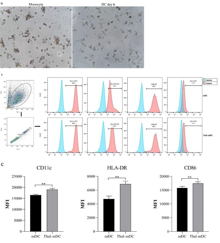
Methods: The immature DCs derived from monocytes were subjected to exposure to Thalidomide and Lipopolysaccharides (LPS) on the fifth day of differentiation, followed by a 24-hour incubation period. On the sixth day, the phenotypic features of the DCs in both the control and treatment groups were assessed using flow cytometry. Subsequently, the gene expression in both the DCs and autologous T cells co-cultured with the DCs was evaluated using the real-time PCR method.
Results: Thalidomide-treated DCs exhibited a significant augmentation in the expression of maturation and stimulatory surface markers CD11c, HLA-DR, and CD86 (P ≤ 0.01), as well as gene expression of TNF-α and IL-12 (P ≤ 0.01) when compared to the control group. Furthermore, co-culture of Thalidomide-treated DCs with T cells increased T-bet and IFN-γ (P ≤ 0.01) expression, while diminished FOXP3 and TGF-β (P ≤ 0.01) expression compared to T cells co-cultured with untreated DCs.
Conclusion: Our findings indicate that in vitro Thalidomide treatment shifts DCs towards an immunogenic state and elevates their T helper 1 inducing capacity, which may be efficient in immunotherapy of various cancers.
BioimpactsPharmacology, Toxicology and Pharmaceutics-Pharmaceutical Science
CiteScore
4.80
自引率
7.70%
发文量
36
审稿时长
5 weeks
期刊介绍:
BioImpacts (BI) is a peer-reviewed multidisciplinary international journal, covering original research articles, reviews, commentaries, hypotheses, methodologies, and visions/reflections dealing with all aspects of biological and biomedical researches at molecular, cellular, functional and translational dimensions.
 求助内容:
求助内容: 应助结果提醒方式:


