Abdullah Merter, Mustafa Özyıldıran, Motohide Shibayama, Zenya Ito, Shu Nakamura, Fujio Ito
{"title":"Comparison of 3 Different Endoscopic Techniques for Lumbar Spinal Stenosis: Comprehensive Radiological and Clinical Study.","authors":"Abdullah Merter, Mustafa Özyıldıran, Motohide Shibayama, Zenya Ito, Shu Nakamura, Fujio Ito","doi":"10.14245/ns.2448864.432","DOIUrl":null,"url":null,"abstract":"<p><strong>Objective: </strong>This study aimed to compare the clinical and comprehensive radiological outcomes of 3 types of endoscopic decompression surgery: unilateral biportal endoscopic lumbar decompression (UBELD), microendoscopic laminotomy (MEL), and percutaneous endoscopic lumbar decompression (PELD).</p><p><strong>Methods: </strong>Patients with single-level lumbar spinal stenosis without instability were included in this multicenter retrospective study. Visual analogue scale (VAS) scores for each extremity, VAS back pain, and Japanese Orthopaedic Association (JOA) scores at preoperative and postoperative 1st, 6th, and 12th months were used as clinical outcome measures. In order to compare the radiological results of the patients, bilateral superior articular distance (SAD), bilateral lateral recess height (LR height), bilateral lateral recess angle (LR angle), and cross-sectional spinal canal area values were measured.</p><p><strong>Results: </strong>Eighty patients in the UBELD group, 73 patients in the MEL group, and 62 patients in the PELD group were included in the study. There was a statistically significant improvement in VAS scores and JOA scores in all groups compared to the preoperative period. At the 12th month postoperatively, the highest lateral decompression values on the approach side were determined as MEL (SAD: 4.1 mm, LR angle: 38.8°, LR height: 4.0 mm), followed by UBELD (SAD: 3.6 mm, LR angle: 36.2°, LR height: 3.3 mm) and PELD (SAD: 3.0 mm, LR angle: 21.7°, LR height: 2.3 mm), respectively. For the contralateral side, the highest lateral recess decompression values were listed as UBELD > MEL > PELD.</p><p><strong>Conclusion: </strong>Effective decompression can be performed using all endoscopic techniques in lumbar spinal stenosis. However lateral recess decompression values were found to be better in UBELD and MEL techniques, compared to PELD.</p>","PeriodicalId":19269,"journal":{"name":"Neurospine","volume":"22 1","pages":"276-285"},"PeriodicalIF":3.6000,"publicationDate":"2025-03-01","publicationTypes":"Journal Article","fieldsOfStudy":null,"isOpenAccess":false,"openAccessPdf":"https://www.ncbi.nlm.nih.gov/pmc/articles/PMC12010855/pdf/","citationCount":"0","resultStr":null,"platform":"Semanticscholar","paperid":null,"PeriodicalName":"Neurospine","FirstCategoryId":"3","ListUrlMain":"https://doi.org/10.14245/ns.2448864.432","RegionNum":2,"RegionCategory":"医学","ArticlePicture":[],"TitleCN":null,"AbstractTextCN":null,"PMCID":null,"EPubDate":"2025/3/31 0:00:00","PubModel":"Epub","JCR":"Q1","JCRName":"CLINICAL NEUROLOGY","Score":null,"Total":0}
引用次数: 0
Abstract
Objective: This study aimed to compare the clinical and comprehensive radiological outcomes of 3 types of endoscopic decompression surgery: unilateral biportal endoscopic lumbar decompression (UBELD), microendoscopic laminotomy (MEL), and percutaneous endoscopic lumbar decompression (PELD).
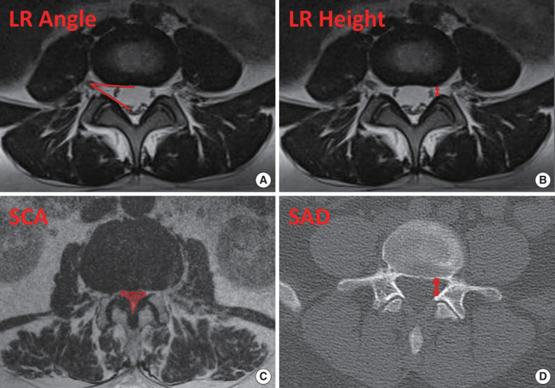
Methods: Patients with single-level lumbar spinal stenosis without instability were included in this multicenter retrospective study. Visual analogue scale (VAS) scores for each extremity, VAS back pain, and Japanese Orthopaedic Association (JOA) scores at preoperative and postoperative 1st, 6th, and 12th months were used as clinical outcome measures. In order to compare the radiological results of the patients, bilateral superior articular distance (SAD), bilateral lateral recess height (LR height), bilateral lateral recess angle (LR angle), and cross-sectional spinal canal area values were measured.
Results: Eighty patients in the UBELD group, 73 patients in the MEL group, and 62 patients in the PELD group were included in the study. There was a statistically significant improvement in VAS scores and JOA scores in all groups compared to the preoperative period. At the 12th month postoperatively, the highest lateral decompression values on the approach side were determined as MEL (SAD: 4.1 mm, LR angle: 38.8°, LR height: 4.0 mm), followed by UBELD (SAD: 3.6 mm, LR angle: 36.2°, LR height: 3.3 mm) and PELD (SAD: 3.0 mm, LR angle: 21.7°, LR height: 2.3 mm), respectively. For the contralateral side, the highest lateral recess decompression values were listed as UBELD > MEL > PELD.
Conclusion: Effective decompression can be performed using all endoscopic techniques in lumbar spinal stenosis. However lateral recess decompression values were found to be better in UBELD and MEL techniques, compared to PELD.
 求助内容:
求助内容: 应助结果提醒方式:


