{"title":"Craniorachischisis Totalis: A Detailed Case Report.","authors":"Sayan Biswas, Praisy Joy, Manisha Gaikwad, Jasmina Begum, Nerbadyswari Deep, Suranjana Banik","doi":"10.22037/ijcn.v19i2.44334","DOIUrl":null,"url":null,"abstract":"<p><p>Neural tube defects (NTDs) are severe congenital anomalies resulting from improper neural tube closure. Craniorachischisis totalis, the most extreme form, involves failure of neural tube formation along the entire cranio-spinal axis. This rare condition is fatal, with limited reported cases globally. We report a case of a 35-year-old G3P1L1A1 woman admitted at 20 weeks and 4 days gestation for medical termination of pregnancy following second-trimester ultrasound findings of anencephaly and spinal dysraphism. The patient began folic acid supplementation only after pregnancy confirmation. The fetus exhibited acrania, bifid vertebrae, exposed neural tissue, frog-eye deformity, and limb contractures. Butterfly vertebrae was observed in infantogram. Retrospective ultrasound review revealed an absent cranial vault, disorganized brain matter, and a large open spinal defect extending to the upper lumbar region. Genetic and infectious panels were largely unremarkable, except for reactive rubella IgG. Craniorachischisis totalis arises from failure of neural tube closure, potentially linked to genetic mutations, folate deficiency, and multiple maternal risk factors. Here, we also revisit the various theories of neural tube closure. Early prenatal diagnosis and counseling are critical for managing craniorachischisis. Periconceptional folic acid supplementation remains the most effective preventive measure.</p>","PeriodicalId":14537,"journal":{"name":"Iranian Journal of Child Neurology","volume":"19 2","pages":"149-153"},"PeriodicalIF":0.9000,"publicationDate":"2025-01-01","publicationTypes":"Journal Article","fieldsOfStudy":null,"isOpenAccess":false,"openAccessPdf":"https://www.ncbi.nlm.nih.gov/pmc/articles/PMC11994127/pdf/","citationCount":"0","resultStr":null,"platform":"Semanticscholar","paperid":null,"PeriodicalName":"Iranian Journal of Child Neurology","FirstCategoryId":"1085","ListUrlMain":"https://doi.org/10.22037/ijcn.v19i2.44334","RegionNum":0,"RegionCategory":null,"ArticlePicture":[],"TitleCN":null,"AbstractTextCN":null,"PMCID":null,"EPubDate":"2025/3/11 0:00:00","PubModel":"Epub","JCR":"Q4","JCRName":"CLINICAL NEUROLOGY","Score":null,"Total":0}
引用次数: 0
Abstract
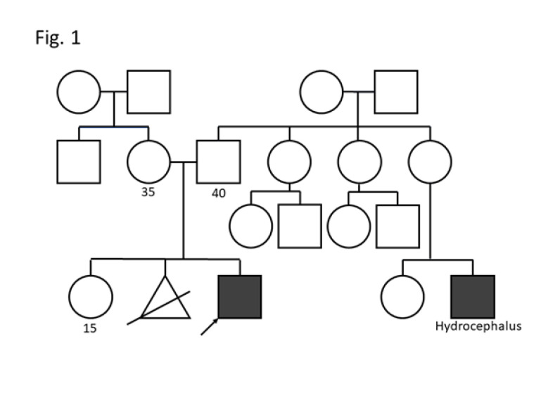
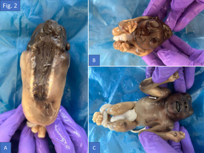
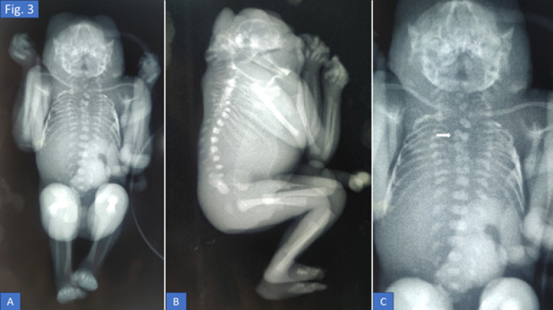
Neural tube defects (NTDs) are severe congenital anomalies resulting from improper neural tube closure. Craniorachischisis totalis, the most extreme form, involves failure of neural tube formation along the entire cranio-spinal axis. This rare condition is fatal, with limited reported cases globally. We report a case of a 35-year-old G3P1L1A1 woman admitted at 20 weeks and 4 days gestation for medical termination of pregnancy following second-trimester ultrasound findings of anencephaly and spinal dysraphism. The patient began folic acid supplementation only after pregnancy confirmation. The fetus exhibited acrania, bifid vertebrae, exposed neural tissue, frog-eye deformity, and limb contractures. Butterfly vertebrae was observed in infantogram. Retrospective ultrasound review revealed an absent cranial vault, disorganized brain matter, and a large open spinal defect extending to the upper lumbar region. Genetic and infectious panels were largely unremarkable, except for reactive rubella IgG. Craniorachischisis totalis arises from failure of neural tube closure, potentially linked to genetic mutations, folate deficiency, and multiple maternal risk factors. Here, we also revisit the various theories of neural tube closure. Early prenatal diagnosis and counseling are critical for managing craniorachischisis. Periconceptional folic acid supplementation remains the most effective preventive measure.

 求助内容:
求助内容: 应助结果提醒方式:


