Dorothy A Thompson, Oliver R Marmoy, Joanne Cowe, Siân E Handley
{"title":"Multi-channel pattern VEPs with full and half field stimulation: methods of interpretation and diagnostic evaluation.","authors":"Dorothy A Thompson, Oliver R Marmoy, Joanne Cowe, Siân E Handley","doi":"10.1007/s10633-025-10012-7","DOIUrl":null,"url":null,"abstract":"<p><strong>Aim: </strong>To describe methods of evaluating multichannel full and half field pattern VEPs using the ISCEV VEP Standard montage.</p><p><strong>Methods: </strong>The dependence of full field and half field pattern VEPs on retinal areas and cortical generators is reviewed and applied to the interpretation and evaluation of multichannel half field pattern VEPs.</p><p><strong>Results: </strong>There are predictable differences in the trans-occipital distributions of components of monocular full, and half field, pattern-reversal and full field, onset-offset VEPs. In combination, the differing distribution and dependence of these components on foveal and macular fields can help to identify and localise chiasmal and retro-chiasmal dysfunction and distinguish this from trans-occipital distribution due to individual variations of cortical architecture. A decision tree synthesising published evidence and current practice is suggested to guide interpretation of trans-occipital VEP distributions.</p><p><strong>Conclusion: </strong>The routine application of two additional lateral channels to acquire multichannel VEPs is quick, easy and adds clinical diagnostic value. The combination of full and half field pattern-reversal and fullfield, onset-offset VEPs can help evaluate chiasmal and retro-chiasmal visual pathway function, and minimise false positive interpretation of asymmetric VEP distributions, which may be due to cortical architecture or cranial anatomy alone.</p>","PeriodicalId":11207,"journal":{"name":"Documenta Ophthalmologica","volume":" ","pages":"87-95"},"PeriodicalIF":2.9000,"publicationDate":"2025-04-01","publicationTypes":"Journal Article","fieldsOfStudy":null,"isOpenAccess":false,"openAccessPdf":"https://www.ncbi.nlm.nih.gov/pmc/articles/PMC11991936/pdf/","citationCount":"0","resultStr":null,"platform":"Semanticscholar","paperid":null,"PeriodicalName":"Documenta Ophthalmologica","FirstCategoryId":"3","ListUrlMain":"https://doi.org/10.1007/s10633-025-10012-7","RegionNum":4,"RegionCategory":"医学","ArticlePicture":[],"TitleCN":null,"AbstractTextCN":null,"PMCID":null,"EPubDate":"2025/3/25 0:00:00","PubModel":"Epub","JCR":"Q2","JCRName":"OPHTHALMOLOGY","Score":null,"Total":0}
引用次数: 0
Abstract
Aim: To describe methods of evaluating multichannel full and half field pattern VEPs using the ISCEV VEP Standard montage.
Methods: The dependence of full field and half field pattern VEPs on retinal areas and cortical generators is reviewed and applied to the interpretation and evaluation of multichannel half field pattern VEPs.
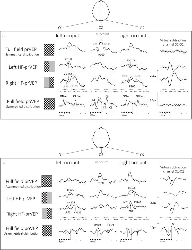
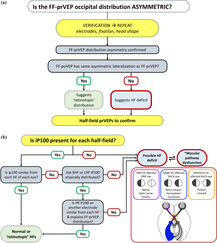
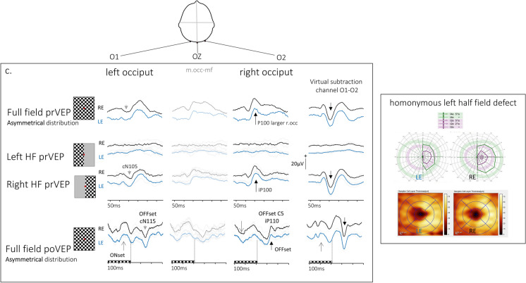
Results: There are predictable differences in the trans-occipital distributions of components of monocular full, and half field, pattern-reversal and full field, onset-offset VEPs. In combination, the differing distribution and dependence of these components on foveal and macular fields can help to identify and localise chiasmal and retro-chiasmal dysfunction and distinguish this from trans-occipital distribution due to individual variations of cortical architecture. A decision tree synthesising published evidence and current practice is suggested to guide interpretation of trans-occipital VEP distributions.
Conclusion: The routine application of two additional lateral channels to acquire multichannel VEPs is quick, easy and adds clinical diagnostic value. The combination of full and half field pattern-reversal and fullfield, onset-offset VEPs can help evaluate chiasmal and retro-chiasmal visual pathway function, and minimise false positive interpretation of asymmetric VEP distributions, which may be due to cortical architecture or cranial anatomy alone.
期刊介绍:
Documenta Ophthalmologica is an official publication of the International Society for Clinical Electrophysiology of Vision. The purpose of the journal is to promote the understanding and application of clinical electrophysiology of vision. Documenta Ophthalmologica will publish reviews, research articles, technical notes, brief reports and case studies which inform the readers about basic and clinical sciences related to visual electrodiagnosis and means to improve diagnosis and clinical management of patients using visual electrophysiology. Studies may involve animals or humans. In either case appropriate care must be taken to follow the Declaration of Helsinki for human subject or appropriate humane standards of animal care (e.g., the ARVO standards on Animal Care and Use).

 求助内容:
求助内容: 应助结果提醒方式:


