The Effectiveness of Endoscopic Ultrasonography and Computed Tomography in the Differentiation of Pancreatic Cystic Neoplasms: A Single-Center Experience.
{"title":"The Effectiveness of Endoscopic Ultrasonography and Computed Tomography in the Differentiation of Pancreatic Cystic Neoplasms: A Single-Center Experience.","authors":"Bengi Öztürk, Koray Ceyhan, Mehmet Bektaş","doi":"10.5152/tjg.2023.23492","DOIUrl":null,"url":null,"abstract":"<p><strong>Background/aims: </strong>Radiological imaging advancements have led to a rise in pancreatic cyst diagnoses. Apart from imaging modalities, endoscopic ultrasonography (EUS) is an important method in the diagnosis of pancreatic cysts. The aim of this study is to determine the clinical, laboratory, biochemical, radiological, and endosonographic features of pancreatic cysts and to assess the effectiveness of EUS and computed tomography (CT) in differentiating between neoplastic and nonneoplastic cysts.</p><p><strong>Materials and methods: </strong>Patients with pancreatic cysts diagnosed by CT, who underwent EUS at the EUS Laboratory of Ankara University Faculty of Medicine, Gastroenterology Department, between 2010 and 2015, were retrospectively evaluated. Size, location, number, and morphological features were compared. Samples for cytology and biochemical tests were obtained through EUS-guided fine needle aspiration (EUS-FNA).</p><p><strong>Results: </strong>The study group included 212 patients. Among them, 125 (59%) patients underwent EUS-FNA. Of these, 63 (52%) were neoplastic, 29 (24%) were nonneoplastic, and 29 (24%) lacked subgroup analysis. The sensitivity of CT in differentiating between neoplastic and nonneoplastic cysts was 82.1% [CI, 68.2%-91.9%], with a specificity of 61.1% (CI, 38.2%-81%) and diagnostic accuracy of 75.4%. Regarding EUS, the sensitivity was 96.7% (CI, 90.2%-99.4%), with a specificity of 45.8% (CI, 27.1%-65.4%) and diagnostic accuracy of 82.3%.</p><p><strong>Conclusion: </strong>Endoscopic ultrasonography demonstrated enhanced sensitivity compared with CT in differentiating neoplastic from nonneoplastic pancreatic cysts. Although no statistical significance was found, this result can be considered clinically remarkable. In addition, EUS displayed distinct advantages in visualizing specific morphological features, emphasizing its potential as a valuable diagnostic tool in assessing pancreatic cystic neoplasms.</p>","PeriodicalId":51205,"journal":{"name":"Turkish Journal of Gastroenterology","volume":"35 12","pages":"945-953"},"PeriodicalIF":1.6000,"publicationDate":"2024-11-28","publicationTypes":"Journal Article","fieldsOfStudy":null,"isOpenAccess":false,"openAccessPdf":"https://www.ncbi.nlm.nih.gov/pmc/articles/PMC11639599/pdf/","citationCount":"0","resultStr":null,"platform":"Semanticscholar","paperid":null,"PeriodicalName":"Turkish Journal of Gastroenterology","FirstCategoryId":"3","ListUrlMain":"https://doi.org/10.5152/tjg.2023.23492","RegionNum":4,"RegionCategory":"医学","ArticlePicture":[],"TitleCN":null,"AbstractTextCN":null,"PMCID":null,"EPubDate":"","PubModel":"","JCR":"Q4","JCRName":"GASTROENTEROLOGY & HEPATOLOGY","Score":null,"Total":0}
引用次数: 0
Abstract
Background/aims: Radiological imaging advancements have led to a rise in pancreatic cyst diagnoses. Apart from imaging modalities, endoscopic ultrasonography (EUS) is an important method in the diagnosis of pancreatic cysts. The aim of this study is to determine the clinical, laboratory, biochemical, radiological, and endosonographic features of pancreatic cysts and to assess the effectiveness of EUS and computed tomography (CT) in differentiating between neoplastic and nonneoplastic cysts.
Materials and methods: Patients with pancreatic cysts diagnosed by CT, who underwent EUS at the EUS Laboratory of Ankara University Faculty of Medicine, Gastroenterology Department, between 2010 and 2015, were retrospectively evaluated. Size, location, number, and morphological features were compared. Samples for cytology and biochemical tests were obtained through EUS-guided fine needle aspiration (EUS-FNA).
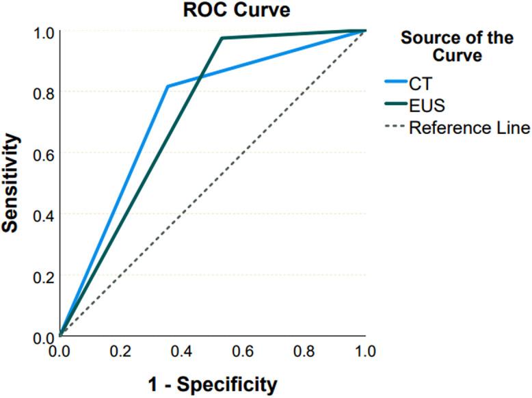
Results: The study group included 212 patients. Among them, 125 (59%) patients underwent EUS-FNA. Of these, 63 (52%) were neoplastic, 29 (24%) were nonneoplastic, and 29 (24%) lacked subgroup analysis. The sensitivity of CT in differentiating between neoplastic and nonneoplastic cysts was 82.1% [CI, 68.2%-91.9%], with a specificity of 61.1% (CI, 38.2%-81%) and diagnostic accuracy of 75.4%. Regarding EUS, the sensitivity was 96.7% (CI, 90.2%-99.4%), with a specificity of 45.8% (CI, 27.1%-65.4%) and diagnostic accuracy of 82.3%.
Conclusion: Endoscopic ultrasonography demonstrated enhanced sensitivity compared with CT in differentiating neoplastic from nonneoplastic pancreatic cysts. Although no statistical significance was found, this result can be considered clinically remarkable. In addition, EUS displayed distinct advantages in visualizing specific morphological features, emphasizing its potential as a valuable diagnostic tool in assessing pancreatic cystic neoplasms.
期刊介绍:
The Turkish Journal of Gastroenterology (Turk J Gastroenterol) is the double-blind peer-reviewed, open access, international publication organ of the Turkish Society of Gastroenterology. The journal is a bimonthly publication, published on January, March, May, July, September, November and its publication language is English.
The Turkish Journal of Gastroenterology aims to publish international at the highest clinical and scientific level on original issues of gastroenterology and hepatology. The journal publishes original papers, review articles, case reports and letters to the editor on clinical and experimental gastroenterology and hepatology.

 求助内容:
求助内容: 应助结果提醒方式:


