Increased deformations are dispensable for encapsulated cell mechanoresponse in engineered bone analogs mimicking aging bone marrow
引用次数: 0
Abstract
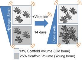
Aged individuals and astronauts experience bone loss despite rigorous physical activity. Bone mechanoresponse is in-part regulated by mesenchymal stem cells (MSCs) that respond to mechanical stimuli. Direct delivery of low intensity vibration (LIV) recovers MSC proliferation in senescence and simulated microgravity models, indicating that age-related reductions in mechanical signal delivery within bone marrow may contribute to declining bone mechanoresponse. To answer this question, we developed a 3D bone marrow analog that controls trabecular geometry, marrow mechanics and external stimuli. Validated finite element (FE) models were developed to quantify strain environment within hydrogels during LIV. Bone marrow analogs with gyroid-based trabeculae of scaffold volume fractions (SV/TV) corresponding to adult (25 %) and aged (13 %) mice were printed using polylactic acid (PLA). MSCs encapsulated in migration-permissive hydrogels within printed trabeculae showed robust cell populations on both PLA surface and hydrogel within a week. Following 14 days of LIV treatment (1 g, 100 Hz, 1 h/day), cell proliferation, type-I collagen (Collagen-I) and filamentous actin (F-actin) were quantified for the cells in the hydrogel fraction. While LIV increased all measured outcomes, FE models predicted higher von Mises strains for the 13 % SV/TV groups (0.2 %) when compared to the 25 % SV/TV group (0.1 %). While LIV increased collagen-I volume 34 % more in 13 % SV/TV groups when compared to 25 % SV/TV groups, collagen-I and F-actin measures remained lower in the 13 % SV/TV groups when compared to 25 % SV/TV counterparts, indicating that both LIV-induced strains and scaffold volume fraction (i.e. available scaffold surface) affect cell behavior in the hydrogel phase. Overall, bone marrow analogs offer a robust and repeatable platform to study bone mechanobiology.

在模仿老化骨髓的工程骨模拟物中,变形的增加对于包裹细胞的机械响应来说是不可或缺的
老年人和宇航员在剧烈运动后仍会出现骨质流失。骨骼的机械反应部分受间充质干细胞(MSCs)的调节,而间充质干细胞会对机械刺激做出反应。在衰老和模拟微重力模型中,低强度振动(LIV)的直接传递可恢复间充质干细胞的增殖,这表明与年龄有关的骨髓内机械信号传递的减少可能会导致骨机械反应的下降。为了回答这个问题,我们开发了一种三维骨髓模拟物,它能控制小梁几何形状、骨髓力学和外部刺激。我们开发了经过验证的有限元(FE)模型,以量化生命周期内水凝胶内的应变环境。使用聚乳酸(PLA)打印了具有陀螺状小梁的骨髓模拟物,其支架体积分数(SV/TV)分别对应成年小鼠(25%)和老年小鼠(13%)。将间叶干细胞包裹在印刷小梁内的迁移许可水凝胶中,一周内聚乳酸表面和水凝胶上都显示出强大的细胞群。经过 14 天的 LIV 处理(1 克、100 赫兹、1 小时/天)后,对水凝胶部分的细胞增殖、I 型胶原蛋白(Collagen-I)和丝状肌动蛋白(F-actin)进行了量化。虽然 LIV 增加了所有测量结果,但与 25% SV/TV 组(0.1%)相比,13% SV/TV 组(0.2%)的 FE 模型预测冯米塞斯应变更高。虽然与 25% SV/TV 组相比,13% SV/TV 组的胶原蛋白-I 体积增加了 34%,但与 25% SV/TV 组相比,13% SV/TV 组的胶原蛋白-I 和 F-肌动蛋白测量值仍然较低,这表明 LIV 诱导的应变和支架体积分数(即可用支架表面)都会影响细胞在水凝胶阶段的行为。总之,骨髓模拟物为研究骨机械生物学提供了一个稳健且可重复的平台。
本文章由计算机程序翻译,如有差异,请以英文原文为准。
求助全文
约1分钟内获得全文
求助全文

 求助内容:
求助内容: 应助结果提醒方式:


