Predicting the clinical prognosis of acute ischemic stroke using machine learning: an application of radiomic biomarkers on non-contrast CT after intravascular interventional treatment.
IF 2.5 4区 医学Q2 MATHEMATICAL & COMPUTATIONAL BIOLOGY
{"title":"Predicting the clinical prognosis of acute ischemic stroke using machine learning: an application of radiomic biomarkers on non-contrast CT after intravascular interventional treatment.","authors":"Hongxian Gu, Yuting Yan, Xiaodong He, Yuyun Xu, Yuguo Wei, Yuan Shao","doi":"10.3389/fninf.2024.1400702","DOIUrl":null,"url":null,"abstract":"<p><strong>Purpose: </strong>This study aimed to develop a radiomic model based on non-contrast computed tomography (NCCT) after interventional treatment to predict the clinical prognosis of acute ischemic stroke (AIS) with large vessel occlusion.</p><p><strong>Methods: </strong>We retrospectively collected 141 cases of AIS from 2016 to 2020 and analyzed the patients' clinical data as well as NCCT data after interventional treatment. Then, the total dataset was divided into training and testing sets according to the subject serial number. The cerebral hemispheres on the infarct side were segmented for radiomics signature extraction. After radiomics signatures were standardized and dimensionality reduced, the training set was used to construct a radiomics model using machine learning. The testing set was then used to validate the prediction model, which was evaluated based on discrimination, calibration, and clinical utility. Finally, a joint model was constructed by incorporating the radiomics signatures and clinical data.</p><p><strong>Results: </strong>The AUCs of the joint model, radiomics signature, NIHSS score, and hypertension were 0.900, 0.863, 0.727, and 0.591, respectively, in the training set. In the testing set, the AUCs of the joint model, radiomics signature, NIHSS score, and hypertension were 0.885, 0.840, 0.721, and 0.590, respectively.</p><p><strong>Conclusion: </strong>Our results provided evidence that using post-interventional NCCT for a radiomic model could be a valuable tool in predicting the clinical prognosis of AIS with large vessel occlusion.</p>","PeriodicalId":12462,"journal":{"name":"Frontiers in Neuroinformatics","volume":"18 ","pages":"1400702"},"PeriodicalIF":2.5000,"publicationDate":"2024-08-22","publicationTypes":"Journal Article","fieldsOfStudy":null,"isOpenAccess":false,"openAccessPdf":"https://www.ncbi.nlm.nih.gov/pmc/articles/PMC11374607/pdf/","citationCount":"0","resultStr":null,"platform":"Semanticscholar","paperid":null,"PeriodicalName":"Frontiers in Neuroinformatics","FirstCategoryId":"3","ListUrlMain":"https://doi.org/10.3389/fninf.2024.1400702","RegionNum":4,"RegionCategory":"医学","ArticlePicture":[],"TitleCN":null,"AbstractTextCN":null,"PMCID":null,"EPubDate":"2024/1/1 0:00:00","PubModel":"eCollection","JCR":"Q2","JCRName":"MATHEMATICAL & COMPUTATIONAL BIOLOGY","Score":null,"Total":0}
引用次数: 0
Abstract
Purpose: This study aimed to develop a radiomic model based on non-contrast computed tomography (NCCT) after interventional treatment to predict the clinical prognosis of acute ischemic stroke (AIS) with large vessel occlusion.
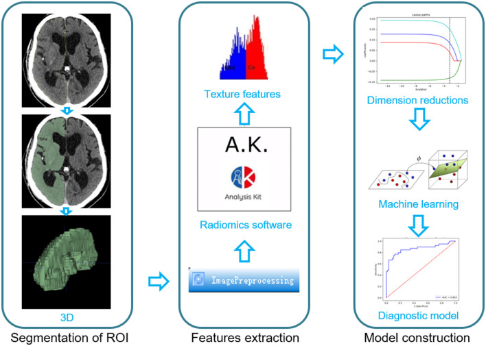
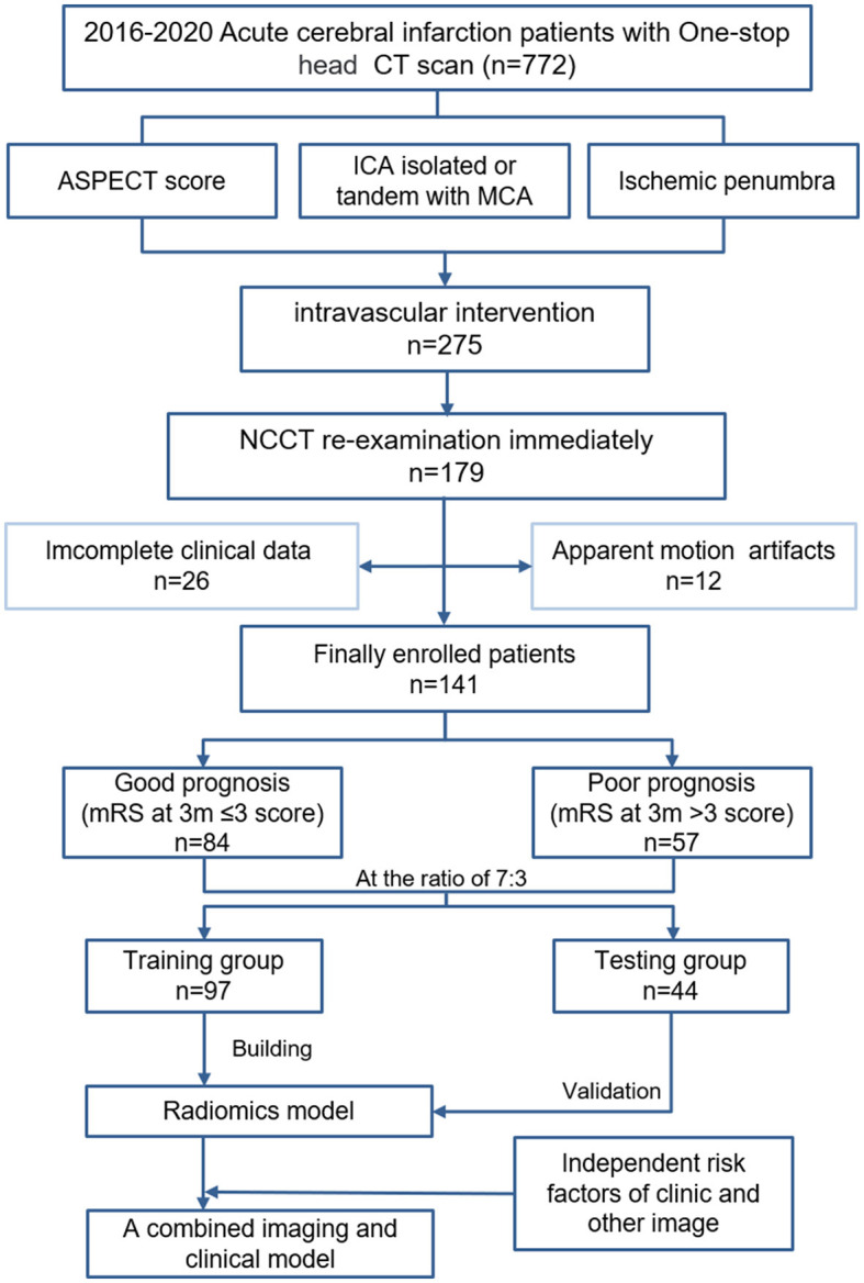
Methods: We retrospectively collected 141 cases of AIS from 2016 to 2020 and analyzed the patients' clinical data as well as NCCT data after interventional treatment. Then, the total dataset was divided into training and testing sets according to the subject serial number. The cerebral hemispheres on the infarct side were segmented for radiomics signature extraction. After radiomics signatures were standardized and dimensionality reduced, the training set was used to construct a radiomics model using machine learning. The testing set was then used to validate the prediction model, which was evaluated based on discrimination, calibration, and clinical utility. Finally, a joint model was constructed by incorporating the radiomics signatures and clinical data.
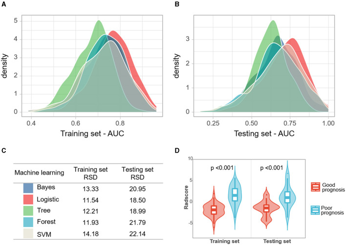
Results: The AUCs of the joint model, radiomics signature, NIHSS score, and hypertension were 0.900, 0.863, 0.727, and 0.591, respectively, in the training set. In the testing set, the AUCs of the joint model, radiomics signature, NIHSS score, and hypertension were 0.885, 0.840, 0.721, and 0.590, respectively.
Conclusion: Our results provided evidence that using post-interventional NCCT for a radiomic model could be a valuable tool in predicting the clinical prognosis of AIS with large vessel occlusion.
期刊介绍:
Frontiers in Neuroinformatics publishes rigorously peer-reviewed research on the development and implementation of numerical/computational models and analytical tools used to share, integrate and analyze experimental data and advance theories of the nervous system functions. Specialty Chief Editors Jan G. Bjaalie at the University of Oslo and Sean L. Hill at the École Polytechnique Fédérale de Lausanne are supported by an outstanding Editorial Board of international experts. This multidisciplinary open-access journal is at the forefront of disseminating and communicating scientific knowledge and impactful discoveries to researchers, academics and the public worldwide.
Neuroscience is being propelled into the information age as the volume of information explodes, demanding organization and synthesis. Novel synthesis approaches are opening up a new dimension for the exploration of the components of brain elements and systems and the vast number of variables that underlie their functions. Neural data is highly heterogeneous with complex inter-relations across multiple levels, driving the need for innovative organizing and synthesizing approaches from genes to cognition, and covering a range of species and disease states.
Frontiers in Neuroinformatics therefore welcomes submissions on existing neuroscience databases, development of data and knowledge bases for all levels of neuroscience, applications and technologies that can facilitate data sharing (interoperability, formats, terminologies, and ontologies), and novel tools for data acquisition, analyses, visualization, and dissemination of nervous system data. Our journal welcomes submissions on new tools (software and hardware) that support brain modeling, and the merging of neuroscience databases with brain models used for simulation and visualization.
 求助内容:
求助内容: 应助结果提醒方式:


