Austin Pereira, Tom Wright, Daniel Weisbrod, Brian G Ballios
{"title":"Vitamin A deficiency retinopathy in the setting of celiac disease and liver fibrosis.","authors":"Austin Pereira, Tom Wright, Daniel Weisbrod, Brian G Ballios","doi":"10.1007/s10633-024-09978-7","DOIUrl":null,"url":null,"abstract":"<p><strong>Purpose: </strong>Vitamin A is a lipid-soluble compound that is critical in maintaining phototransduction. Ocular manifestations of hypovitaminosis A may present with anterior segment signs of xeropthalmia, with advanced cases also causing classic retinal and electrophysiologic changes of vitamin A deficiency retinopathy. We present a case of vitamin A deficiency retinopathy, with corresponding retinal imaging and electrophysiology, in an adult patient with celiac disease and liver fibrosis.</p><p><strong>Methods: </strong>A single case report was conducted in Toronto, Canada.</p><p><strong>Results: </strong>A 77-year-old male with known celiac disease and liver fibrosis presented progressively worsening vision noticed primarily when driving. Vision was 20/50 OD and 20/200 OS. Bitot spots were noted on anterior segment examination. Fundus photography demonstrated bilateral peripheral macular hypopigmentation and far-peripheral granular retinal hypopigmentation with focal yellow dots and hyper-pigmented deposits. Optical coherence tomography (OCT) imaging demonstrated indistinct outer retinal banding with mild outer nuclear layer thinning, focal hyper-reflective deposits, and a thin choroid bilaterally. Full-field electroretinography (ERG) testing demonstrated reduced rod-isolated and combined rod-cone response amplitudes, and multifocal ERG testing demonstrated blunted individual responses throughout the field. The patient was treated with pulse vitamin A therapy. After 6 months of therapy, ERG responses were back within reference range, and the outer retinal changes reversed; visual acuity improved to 20/30 OD and 20/40 OS.</p><p><strong>Conclusion: </strong>This case represents the classic findings of vitamin A deficiency retinopathy on fundus examination and electrophysiologic testing secondary to gastrointestinal pathology. Prompt treatment of high dose vitamin A supplementation led to improvement of full-field and multifocal ERG results, as well as reconstitution of outer retinal architecture.</p>","PeriodicalId":11207,"journal":{"name":"Documenta Ophthalmologica","volume":" ","pages":"125-131"},"PeriodicalIF":2.9000,"publicationDate":"2024-10-01","publicationTypes":"Journal Article","fieldsOfStudy":null,"isOpenAccess":false,"openAccessPdf":"","citationCount":"0","resultStr":null,"platform":"Semanticscholar","paperid":null,"PeriodicalName":"Documenta Ophthalmologica","FirstCategoryId":"3","ListUrlMain":"https://doi.org/10.1007/s10633-024-09978-7","RegionNum":4,"RegionCategory":"医学","ArticlePicture":[],"TitleCN":null,"AbstractTextCN":null,"PMCID":null,"EPubDate":"2024/6/3 0:00:00","PubModel":"Epub","JCR":"Q2","JCRName":"OPHTHALMOLOGY","Score":null,"Total":0}
引用次数: 0
Abstract
Purpose: Vitamin A is a lipid-soluble compound that is critical in maintaining phototransduction. Ocular manifestations of hypovitaminosis A may present with anterior segment signs of xeropthalmia, with advanced cases also causing classic retinal and electrophysiologic changes of vitamin A deficiency retinopathy. We present a case of vitamin A deficiency retinopathy, with corresponding retinal imaging and electrophysiology, in an adult patient with celiac disease and liver fibrosis.
Methods: A single case report was conducted in Toronto, Canada.
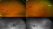
Results: A 77-year-old male with known celiac disease and liver fibrosis presented progressively worsening vision noticed primarily when driving. Vision was 20/50 OD and 20/200 OS. Bitot spots were noted on anterior segment examination. Fundus photography demonstrated bilateral peripheral macular hypopigmentation and far-peripheral granular retinal hypopigmentation with focal yellow dots and hyper-pigmented deposits. Optical coherence tomography (OCT) imaging demonstrated indistinct outer retinal banding with mild outer nuclear layer thinning, focal hyper-reflective deposits, and a thin choroid bilaterally. Full-field electroretinography (ERG) testing demonstrated reduced rod-isolated and combined rod-cone response amplitudes, and multifocal ERG testing demonstrated blunted individual responses throughout the field. The patient was treated with pulse vitamin A therapy. After 6 months of therapy, ERG responses were back within reference range, and the outer retinal changes reversed; visual acuity improved to 20/30 OD and 20/40 OS.
Conclusion: This case represents the classic findings of vitamin A deficiency retinopathy on fundus examination and electrophysiologic testing secondary to gastrointestinal pathology. Prompt treatment of high dose vitamin A supplementation led to improvement of full-field and multifocal ERG results, as well as reconstitution of outer retinal architecture.
目的:维生素 A 是一种脂溶性化合物,对维持光传导至关重要。维生素 A 缺乏症的眼部表现可能表现为前段症状,如干眼症,晚期病例还会引起典型的维生素 A 缺乏性视网膜病变的视网膜和电生理学改变。我们报告了一例患有乳糜泻和肝纤维化的成年患者的维生素 A 缺乏性视网膜病变病例,以及相应的视网膜成像和电生理学病变:方法:在加拿大多伦多进行了单个病例报告:结果:一名 77 岁男性患者,已知患有乳糜泻和肝纤维化,视力逐渐恶化,主要是在开车时。视力为 20/50 OD 和 20/200 OS。前段检查发现有比特斑。眼底照片显示双侧周边黄斑色素减退,远周边颗粒状视网膜色素减退,伴局灶性黄点和色素沉着。光学相干断层扫描(OCT)成像显示,双侧视网膜外带模糊不清,核外层轻度变薄,局灶性高反射沉积,脉络膜变薄。全场视网膜电图(ERG)测试显示,杆隔离和杆-锥联合反应振幅减弱,多灶ERG测试显示,整个视野中的单个反应减弱。患者接受了脉冲维生素 A 治疗。治疗 6 个月后,ERG 反应恢复到参考范围内,视网膜外侧的变化也发生了逆转;视力提高到 20/30 OD 和 20/40 OS:本病例是继发于胃肠道病变的维生素 A 缺乏性视网膜病变的典型眼底检查和电生理测试结果。及时补充大剂量维生素 A 可改善全视野和多焦点 ERG 结果,并重建外层视网膜结构。
期刊介绍:
Documenta Ophthalmologica is an official publication of the International Society for Clinical Electrophysiology of Vision. The purpose of the journal is to promote the understanding and application of clinical electrophysiology of vision. Documenta Ophthalmologica will publish reviews, research articles, technical notes, brief reports and case studies which inform the readers about basic and clinical sciences related to visual electrodiagnosis and means to improve diagnosis and clinical management of patients using visual electrophysiology. Studies may involve animals or humans. In either case appropriate care must be taken to follow the Declaration of Helsinki for human subject or appropriate humane standards of animal care (e.g., the ARVO standards on Animal Care and Use).

 求助内容:
求助内容: 应助结果提醒方式:


