{"title":"Current Perspectives on Type 3 Macular Neovascularization due to Age-Related Macular Degeneration.","authors":"Christoph R Clemens, Nicole Eter, Florian Alten","doi":"10.1159/000536278","DOIUrl":null,"url":null,"abstract":"<p><strong>Background: </strong>The aim of this review was to systematically summarize the current knowledge on type 3 macular neovascularization (MNV3) in age-related macular degeneration (AMD).</p><p><strong>Summary: </strong>Recent histopathologic and multimodal imaging findings led to the consensus definition of the new term \"type 3 macular neovascularization\" in AMD. MNV3 originates in the deep vascular plexus as a neovascular process without connection with the retinal pigment epithelium in the initial stages. This type has numerous clinical and pathomorphologic features that separate it from the other two types of MNV in AMD. Besides, its frequency appears to be higher than previously thought. In optical coherence tomography (OCT), MNV3 can be classified into stages 1-3. Hyperreflective foci in the outer retina possibly represent a precursor lesion. In addition, MNV3 is characterized by a strong association with reticular pseudodrusen, a high rate of bilaterality, close associations with advanced age and arterial hypertension, decreased choroidal thickness, and decreased choriocapillaris flow signals. Data from latest anti-vascular endothelial growth factor studies in MNV3 suggest that the OCT biomarkers in intraretinal and subretinal fluids should be interpreted differently than in the other types. Additionally, data from MNV3 eyes should be analyzed separately, allowing optimal type-specific treatment strategies in the future.</p><p><strong>Key messages: </strong>This review highlights the need for accurate characterization of neovascular AMD lesions and an MNV type-specific approach, particularly for MNV3.</p>","PeriodicalId":19595,"journal":{"name":"Ophthalmologica","volume":" ","pages":"73-84"},"PeriodicalIF":1.9000,"publicationDate":"2024-01-01","publicationTypes":"Journal Article","fieldsOfStudy":null,"isOpenAccess":false,"openAccessPdf":"https://www.ncbi.nlm.nih.gov/pmc/articles/PMC11160428/pdf/","citationCount":"0","resultStr":null,"platform":"Semanticscholar","paperid":null,"PeriodicalName":"Ophthalmologica","FirstCategoryId":"3","ListUrlMain":"https://doi.org/10.1159/000536278","RegionNum":4,"RegionCategory":"医学","ArticlePicture":[],"TitleCN":null,"AbstractTextCN":null,"PMCID":null,"EPubDate":"2024/1/24 0:00:00","PubModel":"Epub","JCR":"Q2","JCRName":"OPHTHALMOLOGY","Score":null,"Total":0}
引用次数: 0
Abstract
Background: The aim of this review was to systematically summarize the current knowledge on type 3 macular neovascularization (MNV3) in age-related macular degeneration (AMD).
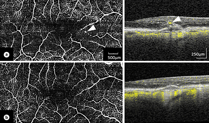
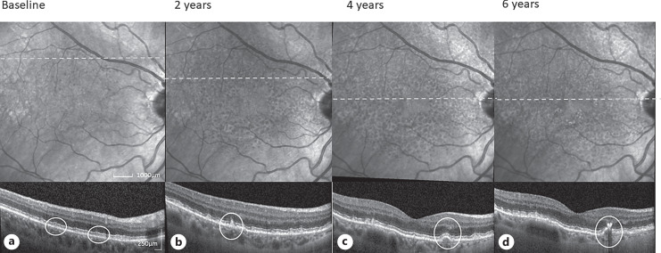
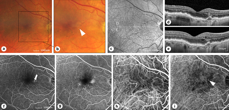
Summary: Recent histopathologic and multimodal imaging findings led to the consensus definition of the new term "type 3 macular neovascularization" in AMD. MNV3 originates in the deep vascular plexus as a neovascular process without connection with the retinal pigment epithelium in the initial stages. This type has numerous clinical and pathomorphologic features that separate it from the other two types of MNV in AMD. Besides, its frequency appears to be higher than previously thought. In optical coherence tomography (OCT), MNV3 can be classified into stages 1-3. Hyperreflective foci in the outer retina possibly represent a precursor lesion. In addition, MNV3 is characterized by a strong association with reticular pseudodrusen, a high rate of bilaterality, close associations with advanced age and arterial hypertension, decreased choroidal thickness, and decreased choriocapillaris flow signals. Data from latest anti-vascular endothelial growth factor studies in MNV3 suggest that the OCT biomarkers in intraretinal and subretinal fluids should be interpreted differently than in the other types. Additionally, data from MNV3 eyes should be analyzed separately, allowing optimal type-specific treatment strategies in the future.
Key messages: This review highlights the need for accurate characterization of neovascular AMD lesions and an MNV type-specific approach, particularly for MNV3.
期刊介绍:
Published since 1899, ''Ophthalmologica'' has become a frequently cited guide to international work in clinical and experimental ophthalmology. It contains a selection of patient-oriented contributions covering the etiology of eye diseases, diagnostic techniques, and advances in medical and surgical treatment. Straightforward, factual reporting provides both interesting and useful reading. In addition to original papers, ''Ophthalmologica'' features regularly timely reviews in an effort to keep the reader well informed and updated. The large international circulation of this journal reflects its importance.
 求助内容:
求助内容: 应助结果提醒方式:


