Maria Vittoria Cicinelli , Prithvi Ramtohul , Alessandro Marchese , Francesco Bandello , K. Bailey Freund , Elisabetta Miserocchi , Lee M. Jampol
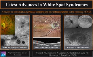
{"title":"Latest advances in white spot syndromes: New findings and interpretations","authors":"Maria Vittoria Cicinelli , Prithvi Ramtohul , Alessandro Marchese , Francesco Bandello , K. Bailey Freund , Elisabetta Miserocchi , Lee M. Jampol","doi":"10.1016/j.preteyeres.2023.101207","DOIUrl":null,"url":null,"abstract":"<div><p>White spot syndromes<span><span><span><span> (WSS) pose challenges in the field of ophthalmology, particularly in terms of accurate diagnosis and effective management. However, recent advancements in </span>multimodal imaging<span> (MMI) have significantly contributed to our understanding of WSS, allowing for improved characterization of these inflammatory chorioretinopathies. By employing various imaging modalities, including fundus fluorescein </span></span>angiography<span>, indocyanine green angiography, fundus autofluorescence, </span></span>optical coherence tomography<span> (OCT), ultra-widefield imaging, and OCT angiography, researchers and clinicians have gained valuable insights into the underlying pathophysiological changes and clinical progression of WSS.</span></span></p><p>Furthermore, MMI has unveiled novel and atypical variants within the spectrum of WSS, expanding our knowledge in this field. Notably, the identification of secondary forms of WSS occurring concurrently with unrelated chorioretinal disorders has suggested a potential autoimmune mechanism underlying these conditions. The introduction of MMI has also facilitated a more comprehensive evaluation of previously ill-defined entities, such as acute zonal occult outer retinopathy, leading to improved diagnostic criteria and enhanced recognition of distinct features. This review paper provides a comprehensive overview of the latest advances and interpretations in WSS. By integrating MMI into the diagnosis and management of these conditions, this review aims to enhance patient outcomes and provide valuable insights into the complexities surrounding WSS.</p></div>","PeriodicalId":21159,"journal":{"name":"Progress in Retinal and Eye Research","volume":"97 ","pages":"Article 101207"},"PeriodicalIF":18.6000,"publicationDate":"2023-08-12","publicationTypes":"Journal Article","fieldsOfStudy":null,"isOpenAccess":false,"openAccessPdf":"","citationCount":"2","resultStr":null,"platform":"Semanticscholar","paperid":null,"PeriodicalName":"Progress in Retinal and Eye Research","FirstCategoryId":"3","ListUrlMain":"https://www.sciencedirect.com/science/article/pii/S1350946223000460","RegionNum":1,"RegionCategory":"医学","ArticlePicture":[],"TitleCN":null,"AbstractTextCN":null,"PMCID":null,"EPubDate":"","PubModel":"","JCR":"Q1","JCRName":"OPHTHALMOLOGY","Score":null,"Total":0}
引用次数: 2
Abstract
White spot syndromes (WSS) pose challenges in the field of ophthalmology, particularly in terms of accurate diagnosis and effective management. However, recent advancements in multimodal imaging (MMI) have significantly contributed to our understanding of WSS, allowing for improved characterization of these inflammatory chorioretinopathies. By employing various imaging modalities, including fundus fluorescein angiography, indocyanine green angiography, fundus autofluorescence, optical coherence tomography (OCT), ultra-widefield imaging, and OCT angiography, researchers and clinicians have gained valuable insights into the underlying pathophysiological changes and clinical progression of WSS.
Furthermore, MMI has unveiled novel and atypical variants within the spectrum of WSS, expanding our knowledge in this field. Notably, the identification of secondary forms of WSS occurring concurrently with unrelated chorioretinal disorders has suggested a potential autoimmune mechanism underlying these conditions. The introduction of MMI has also facilitated a more comprehensive evaluation of previously ill-defined entities, such as acute zonal occult outer retinopathy, leading to improved diagnostic criteria and enhanced recognition of distinct features. This review paper provides a comprehensive overview of the latest advances and interpretations in WSS. By integrating MMI into the diagnosis and management of these conditions, this review aims to enhance patient outcomes and provide valuable insights into the complexities surrounding WSS.
期刊介绍:
Progress in Retinal and Eye Research is a Reviews-only journal. By invitation, leading experts write on basic and clinical aspects of the eye in a style appealing to molecular biologists, neuroscientists and physiologists, as well as to vision researchers and ophthalmologists.
The journal covers all aspects of eye research, including topics pertaining to the retina and pigment epithelial layer, cornea, tears, lacrimal glands, aqueous humour, iris, ciliary body, trabeculum, lens, vitreous humour and diseases such as dry-eye, inflammation, keratoconus, corneal dystrophy, glaucoma and cataract.
 求助内容:
求助内容: 应助结果提醒方式:


