{"title":"低真空扫描电镜对环氧树脂块肾活检的直接观察。","authors":"Akihiro Tojo, Makoto Abe, Kin-Ichi Matsuyama","doi":"10.1007/s00795-023-00356-x","DOIUrl":null,"url":null,"abstract":"<p><p>To improve the resolution of low-vacuum scanning electron microscopy (LVSEM), the epoxy resin block for the transmission electron microscopy (TEM) was observed directly with LVSEM. After observing ultrathin sections from renal biopsies of IgA nephropathy, membranous nephropathy, lupus nephritis, diabetic nephropathy (DM), thin basement membrane disease (TBMD), Alport's syndrome, Fabry's disease, and renal amyloidosis, the epoxy resin blocks of the same sites were observed by LVSEM and compared. The LVSEM image of the epoxy resin block corresponds to the negative of the TEM image, and when the gradation is reversed, the LVSEM image was comparable to the TEM image. At a low magnification of 100 ×, the entire specimen, including the glomerulus, was obtained. LVSEM at 5000 × magnification was sufficient to identify paramesangial deposits in IgA nephropathy and subepithelial electron-dense deposits (EDD) and spikes in membranous nephropathy. Glomerular basement membrane thickening in DM and thinning in TBMD could be sufficiently diagnosed with LVSEM at 6000 ×. Accumulation of ceramide in Fabry's disease was easily identified, but amyloid fibril could not be identified by LVSEM. LVSEM of renal biopsy epoxy resin blocks can replace TEM up to moderate magnification.</p>","PeriodicalId":18338,"journal":{"name":"Medical Molecular Morphology","volume":"56 3","pages":"206-216"},"PeriodicalIF":1.1000,"publicationDate":"2023-09-01","publicationTypes":"Journal Article","fieldsOfStudy":null,"isOpenAccess":false,"openAccessPdf":"https://www.ncbi.nlm.nih.gov/pmc/articles/PMC10415507/pdf/","citationCount":"0","resultStr":"{\"title\":\"Direct observation of epoxy resin blocks for renal biopsy by low-vacuum scanning electron microscopy.\",\"authors\":\"Akihiro Tojo, Makoto Abe, Kin-Ichi Matsuyama\",\"doi\":\"10.1007/s00795-023-00356-x\",\"DOIUrl\":null,\"url\":null,\"abstract\":\"<p><p>To improve the resolution of low-vacuum scanning electron microscopy (LVSEM), the epoxy resin block for the transmission electron microscopy (TEM) was observed directly with LVSEM. After observing ultrathin sections from renal biopsies of IgA nephropathy, membranous nephropathy, lupus nephritis, diabetic nephropathy (DM), thin basement membrane disease (TBMD), Alport's syndrome, Fabry's disease, and renal amyloidosis, the epoxy resin blocks of the same sites were observed by LVSEM and compared. The LVSEM image of the epoxy resin block corresponds to the negative of the TEM image, and when the gradation is reversed, the LVSEM image was comparable to the TEM image. At a low magnification of 100 ×, the entire specimen, including the glomerulus, was obtained. LVSEM at 5000 × magnification was sufficient to identify paramesangial deposits in IgA nephropathy and subepithelial electron-dense deposits (EDD) and spikes in membranous nephropathy. Glomerular basement membrane thickening in DM and thinning in TBMD could be sufficiently diagnosed with LVSEM at 6000 ×. Accumulation of ceramide in Fabry's disease was easily identified, but amyloid fibril could not be identified by LVSEM. LVSEM of renal biopsy epoxy resin blocks can replace TEM up to moderate magnification.</p>\",\"PeriodicalId\":18338,\"journal\":{\"name\":\"Medical Molecular Morphology\",\"volume\":\"56 3\",\"pages\":\"206-216\"},\"PeriodicalIF\":1.1000,\"publicationDate\":\"2023-09-01\",\"publicationTypes\":\"Journal Article\",\"fieldsOfStudy\":null,\"isOpenAccess\":false,\"openAccessPdf\":\"https://www.ncbi.nlm.nih.gov/pmc/articles/PMC10415507/pdf/\",\"citationCount\":\"0\",\"resultStr\":null,\"platform\":\"Semanticscholar\",\"paperid\":null,\"PeriodicalName\":\"Medical Molecular Morphology\",\"FirstCategoryId\":\"3\",\"ListUrlMain\":\"https://doi.org/10.1007/s00795-023-00356-x\",\"RegionNum\":4,\"RegionCategory\":\"医学\",\"ArticlePicture\":[],\"TitleCN\":null,\"AbstractTextCN\":null,\"PMCID\":null,\"EPubDate\":\"2023/5/10 0:00:00\",\"PubModel\":\"Epub\",\"JCR\":\"Q3\",\"JCRName\":\"PATHOLOGY\",\"Score\":null,\"Total\":0}","platform":"Semanticscholar","paperid":null,"PeriodicalName":"Medical Molecular Morphology","FirstCategoryId":"3","ListUrlMain":"https://doi.org/10.1007/s00795-023-00356-x","RegionNum":4,"RegionCategory":"医学","ArticlePicture":[],"TitleCN":null,"AbstractTextCN":null,"PMCID":null,"EPubDate":"2023/5/10 0:00:00","PubModel":"Epub","JCR":"Q3","JCRName":"PATHOLOGY","Score":null,"Total":0}
Direct observation of epoxy resin blocks for renal biopsy by low-vacuum scanning electron microscopy.
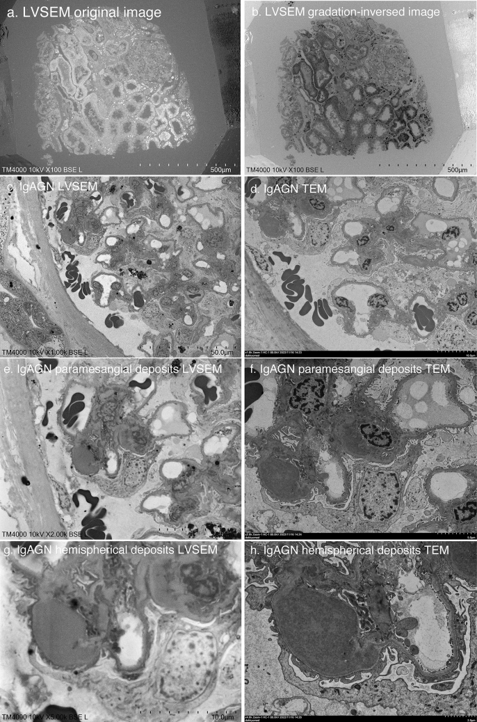
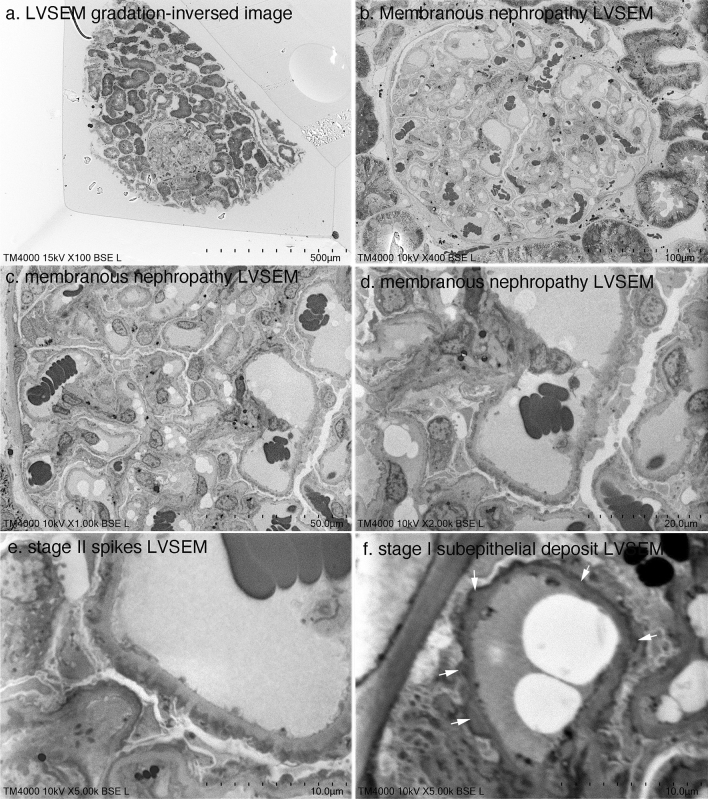
To improve the resolution of low-vacuum scanning electron microscopy (LVSEM), the epoxy resin block for the transmission electron microscopy (TEM) was observed directly with LVSEM. After observing ultrathin sections from renal biopsies of IgA nephropathy, membranous nephropathy, lupus nephritis, diabetic nephropathy (DM), thin basement membrane disease (TBMD), Alport's syndrome, Fabry's disease, and renal amyloidosis, the epoxy resin blocks of the same sites were observed by LVSEM and compared. The LVSEM image of the epoxy resin block corresponds to the negative of the TEM image, and when the gradation is reversed, the LVSEM image was comparable to the TEM image. At a low magnification of 100 ×, the entire specimen, including the glomerulus, was obtained. LVSEM at 5000 × magnification was sufficient to identify paramesangial deposits in IgA nephropathy and subepithelial electron-dense deposits (EDD) and spikes in membranous nephropathy. Glomerular basement membrane thickening in DM and thinning in TBMD could be sufficiently diagnosed with LVSEM at 6000 ×. Accumulation of ceramide in Fabry's disease was easily identified, but amyloid fibril could not be identified by LVSEM. LVSEM of renal biopsy epoxy resin blocks can replace TEM up to moderate magnification.
期刊介绍:
Medical Molecular Morphology is an international forum for researchers in both basic and clinical medicine to present and discuss new research on the structural mechanisms and the processes of health and disease at the molecular level. The structures of molecules, organelles, cells, tissues, and organs determine their normal function. Disease is thus best understood in terms of structural changes in these different levels of biological organization, especially in molecules and molecular interactions as well as the cellular localization of chemical components. Medical Molecular Morphology welcomes articles on basic or clinical research in the fields of cell biology, molecular biology, and medical, veterinary, and dental sciences using techniques for structural research such as electron microscopy, confocal laser scanning microscopy, enzyme histochemistry, immunohistochemistry, radioautography, X-ray microanalysis, and in situ hybridization.
Manuscripts submitted for publication must contain a statement to the effect that all human studies have been reviewed by the appropriate ethics committee and have therefore been performed in accordance with the ethical standards laid down in an appropriate version of the 1964 Declaration of Helsinki. It should also be stated clearly in the text that all persons gave their informed consent prior to their inclusion in the study. Details that might disclose the identity of the subjects under study should be omitted.
 求助内容:
求助内容: 应助结果提醒方式:


