{"title":"盆底和会阴肌:骨骼肌和平滑肌在盆底稳定中的动态协调","authors":"Satoru Muro, Keiichi Akita","doi":"10.1007/s12565-023-00717-7","DOIUrl":null,"url":null,"abstract":"<div><p>The purpose of this review is to present our researches on the pelvic outlet muscles, including the pelvic floor and perineal muscles, which are responsible for urinary function, defecation, sexual function, and core stability, and to discuss the insights into the mechanism of pelvic floor stabilization based on the findings. Our studies are conducted using a combination of macroscopic examination, immunohistological analysis, 3D reconstruction, and imaging. Unlike most previous reports, this article describes not only on skeletal muscle but also on smooth muscle structures in the pelvic floor and perineum to encourage new understanding. The skeletal muscles of the pelvic outlet are continuous, which means that they share muscle bundles. They form three muscle slings that pass anterior and posterior to the anal canal, thus serving as the foundation of pelvic floor support. The smooth muscle of the pelvic outlet, in addition to forming the walls of the viscera, also extends in three dimensions. This continuous smooth muscle occupies the central region of the pelvic floor and perineum, thus revising the conventional understanding of the perineal body. At the interface between the levator ani and pelvic viscera, smooth muscle forms characteristic structures that transfer the lifting power of the levator ani to the pelvic viscera. The findings suggest new concepts of pelvic floor stabilization mechanisms, such as dynamic coordination between skeletal and smooth muscles. These two types of muscles possibly coordinate the direction and force of muscle contraction with each other.</p></div>","PeriodicalId":7816,"journal":{"name":"Anatomical Science International","volume":"98 3","pages":"407 - 425"},"PeriodicalIF":1.2000,"publicationDate":"2023-03-24","publicationTypes":"Journal Article","fieldsOfStudy":null,"isOpenAccess":false,"openAccessPdf":"https://link.springer.com/content/pdf/10.1007/s12565-023-00717-7.pdf","citationCount":"2","resultStr":"{\"title\":\"Pelvic floor and perineal muscles: a dynamic coordination between skeletal and smooth muscles on pelvic floor stabilization\",\"authors\":\"Satoru Muro, Keiichi Akita\",\"doi\":\"10.1007/s12565-023-00717-7\",\"DOIUrl\":null,\"url\":null,\"abstract\":\"<div><p>The purpose of this review is to present our researches on the pelvic outlet muscles, including the pelvic floor and perineal muscles, which are responsible for urinary function, defecation, sexual function, and core stability, and to discuss the insights into the mechanism of pelvic floor stabilization based on the findings. Our studies are conducted using a combination of macroscopic examination, immunohistological analysis, 3D reconstruction, and imaging. Unlike most previous reports, this article describes not only on skeletal muscle but also on smooth muscle structures in the pelvic floor and perineum to encourage new understanding. The skeletal muscles of the pelvic outlet are continuous, which means that they share muscle bundles. They form three muscle slings that pass anterior and posterior to the anal canal, thus serving as the foundation of pelvic floor support. The smooth muscle of the pelvic outlet, in addition to forming the walls of the viscera, also extends in three dimensions. This continuous smooth muscle occupies the central region of the pelvic floor and perineum, thus revising the conventional understanding of the perineal body. At the interface between the levator ani and pelvic viscera, smooth muscle forms characteristic structures that transfer the lifting power of the levator ani to the pelvic viscera. The findings suggest new concepts of pelvic floor stabilization mechanisms, such as dynamic coordination between skeletal and smooth muscles. These two types of muscles possibly coordinate the direction and force of muscle contraction with each other.</p></div>\",\"PeriodicalId\":7816,\"journal\":{\"name\":\"Anatomical Science International\",\"volume\":\"98 3\",\"pages\":\"407 - 425\"},\"PeriodicalIF\":1.2000,\"publicationDate\":\"2023-03-24\",\"publicationTypes\":\"Journal Article\",\"fieldsOfStudy\":null,\"isOpenAccess\":false,\"openAccessPdf\":\"https://link.springer.com/content/pdf/10.1007/s12565-023-00717-7.pdf\",\"citationCount\":\"2\",\"resultStr\":null,\"platform\":\"Semanticscholar\",\"paperid\":null,\"PeriodicalName\":\"Anatomical Science International\",\"FirstCategoryId\":\"3\",\"ListUrlMain\":\"https://link.springer.com/article/10.1007/s12565-023-00717-7\",\"RegionNum\":4,\"RegionCategory\":\"医学\",\"ArticlePicture\":[],\"TitleCN\":null,\"AbstractTextCN\":null,\"PMCID\":null,\"EPubDate\":\"\",\"PubModel\":\"\",\"JCR\":\"Q3\",\"JCRName\":\"ANATOMY & MORPHOLOGY\",\"Score\":null,\"Total\":0}","platform":"Semanticscholar","paperid":null,"PeriodicalName":"Anatomical Science International","FirstCategoryId":"3","ListUrlMain":"https://link.springer.com/article/10.1007/s12565-023-00717-7","RegionNum":4,"RegionCategory":"医学","ArticlePicture":[],"TitleCN":null,"AbstractTextCN":null,"PMCID":null,"EPubDate":"","PubModel":"","JCR":"Q3","JCRName":"ANATOMY & MORPHOLOGY","Score":null,"Total":0}
Pelvic floor and perineal muscles: a dynamic coordination between skeletal and smooth muscles on pelvic floor stabilization
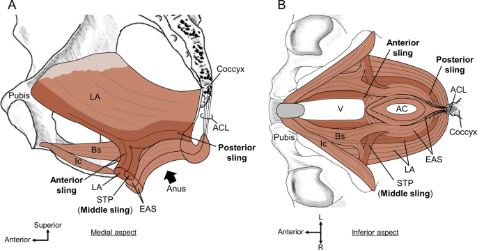
The purpose of this review is to present our researches on the pelvic outlet muscles, including the pelvic floor and perineal muscles, which are responsible for urinary function, defecation, sexual function, and core stability, and to discuss the insights into the mechanism of pelvic floor stabilization based on the findings. Our studies are conducted using a combination of macroscopic examination, immunohistological analysis, 3D reconstruction, and imaging. Unlike most previous reports, this article describes not only on skeletal muscle but also on smooth muscle structures in the pelvic floor and perineum to encourage new understanding. The skeletal muscles of the pelvic outlet are continuous, which means that they share muscle bundles. They form three muscle slings that pass anterior and posterior to the anal canal, thus serving as the foundation of pelvic floor support. The smooth muscle of the pelvic outlet, in addition to forming the walls of the viscera, also extends in three dimensions. This continuous smooth muscle occupies the central region of the pelvic floor and perineum, thus revising the conventional understanding of the perineal body. At the interface between the levator ani and pelvic viscera, smooth muscle forms characteristic structures that transfer the lifting power of the levator ani to the pelvic viscera. The findings suggest new concepts of pelvic floor stabilization mechanisms, such as dynamic coordination between skeletal and smooth muscles. These two types of muscles possibly coordinate the direction and force of muscle contraction with each other.
期刊介绍:
The official English journal of the Japanese Association of Anatomists, Anatomical Science International (formerly titled Kaibogaku Zasshi) publishes original research articles dealing with morphological sciences.
Coverage in the journal includes molecular, cellular, histological and gross anatomical studies on humans and on normal and experimental animals, as well as functional morphological, biochemical, physiological and behavioral studies if they include morphological analysis.

 求助内容:
求助内容: 应助结果提醒方式:


