Jasmin Kniese , Sven Ritschar , Lina Bünger , Heike Feldhaar , Christian Laforsch , Andreas Römpp , Heinar Schmidt
{"title":"利用拉曼光谱成像定位和鉴定组织切片中的聚苯乙烯颗粒","authors":"Jasmin Kniese , Sven Ritschar , Lina Bünger , Heike Feldhaar , Christian Laforsch , Andreas Römpp , Heinar Schmidt","doi":"10.1016/j.impact.2023.100465","DOIUrl":null,"url":null,"abstract":"<div><p>The uptake of microplastic particles (MPP) by organisms is frequently described and poses a potential risk for these organisms and ultimately for humans either through direct uptake or trophic transfer. Currently, the in-situ detection of MPP in organisms is typically based on histological examination of tissue sections after uptake of fluorescently-labelled MPP and is thus not feasible for environmental samples. The alternative approach is purification of MPP from whole organisms or organs by chemical digestion and subsequent spectroscopic detection (FT-IR or Raman). While this approach is feasible for un-labelled particles it goes along with loss of any spatial information related to the location in the tissue. In our study we aimed at providing a workflow for the localisation and identification of non-fluorescent and fluorescent polystyrene (PS) particles (fragments, size range 2–130 μm) in tissue sections of the model organism <em>Eisenia fetida</em> with Raman spectroscopic imaging (RSI). We provide methodological approaches for the preparation of the samples, technical parameters for the RSI measurements and data analysis for PS differentiation in tissue sections. The developed approaches were combined in a workflow for the in-situ analysis of MPP in tissue sections. The spectroscopic analysis requires differentiation of spectra of MPP and interfering compounds, which is challenging given the complexity of tissue. Therefore, a classification algorithm was developed to differentiate PS particles from haem, intestinal contents and surrounding tissue. It allows the differentiation of PS particles from protein in the tissue of <em>E. fetida</em> with an accuracy of 95%. The smallest PS particle detected in the tissue was 2 μm in diameter. We show that it is possible to localise and identify non-fluorescent and fluorescent ingested PS particles directly in tissue sections of <em>E. fetida</em> in the gut lumen and the adjacent tissue.</p></div>","PeriodicalId":18786,"journal":{"name":"NanoImpact","volume":"30 ","pages":"Article 100465"},"PeriodicalIF":4.7000,"publicationDate":"2023-04-01","publicationTypes":"Journal Article","fieldsOfStudy":null,"isOpenAccess":false,"openAccessPdf":"","citationCount":"1","resultStr":"{\"title\":\"Localisation and identification of polystyrene particles in tissue sections using Raman spectroscopic imaging\",\"authors\":\"Jasmin Kniese , Sven Ritschar , Lina Bünger , Heike Feldhaar , Christian Laforsch , Andreas Römpp , Heinar Schmidt\",\"doi\":\"10.1016/j.impact.2023.100465\",\"DOIUrl\":null,\"url\":null,\"abstract\":\"<div><p>The uptake of microplastic particles (MPP) by organisms is frequently described and poses a potential risk for these organisms and ultimately for humans either through direct uptake or trophic transfer. Currently, the in-situ detection of MPP in organisms is typically based on histological examination of tissue sections after uptake of fluorescently-labelled MPP and is thus not feasible for environmental samples. The alternative approach is purification of MPP from whole organisms or organs by chemical digestion and subsequent spectroscopic detection (FT-IR or Raman). While this approach is feasible for un-labelled particles it goes along with loss of any spatial information related to the location in the tissue. In our study we aimed at providing a workflow for the localisation and identification of non-fluorescent and fluorescent polystyrene (PS) particles (fragments, size range 2–130 μm) in tissue sections of the model organism <em>Eisenia fetida</em> with Raman spectroscopic imaging (RSI). We provide methodological approaches for the preparation of the samples, technical parameters for the RSI measurements and data analysis for PS differentiation in tissue sections. The developed approaches were combined in a workflow for the in-situ analysis of MPP in tissue sections. The spectroscopic analysis requires differentiation of spectra of MPP and interfering compounds, which is challenging given the complexity of tissue. Therefore, a classification algorithm was developed to differentiate PS particles from haem, intestinal contents and surrounding tissue. It allows the differentiation of PS particles from protein in the tissue of <em>E. fetida</em> with an accuracy of 95%. The smallest PS particle detected in the tissue was 2 μm in diameter. We show that it is possible to localise and identify non-fluorescent and fluorescent ingested PS particles directly in tissue sections of <em>E. fetida</em> in the gut lumen and the adjacent tissue.</p></div>\",\"PeriodicalId\":18786,\"journal\":{\"name\":\"NanoImpact\",\"volume\":\"30 \",\"pages\":\"Article 100465\"},\"PeriodicalIF\":4.7000,\"publicationDate\":\"2023-04-01\",\"publicationTypes\":\"Journal Article\",\"fieldsOfStudy\":null,\"isOpenAccess\":false,\"openAccessPdf\":\"\",\"citationCount\":\"1\",\"resultStr\":null,\"platform\":\"Semanticscholar\",\"paperid\":null,\"PeriodicalName\":\"NanoImpact\",\"FirstCategoryId\":\"93\",\"ListUrlMain\":\"https://www.sciencedirect.com/science/article/pii/S2452074823000162\",\"RegionNum\":3,\"RegionCategory\":\"环境科学与生态学\",\"ArticlePicture\":[],\"TitleCN\":null,\"AbstractTextCN\":null,\"PMCID\":null,\"EPubDate\":\"\",\"PubModel\":\"\",\"JCR\":\"Q2\",\"JCRName\":\"ENVIRONMENTAL SCIENCES\",\"Score\":null,\"Total\":0}","platform":"Semanticscholar","paperid":null,"PeriodicalName":"NanoImpact","FirstCategoryId":"93","ListUrlMain":"https://www.sciencedirect.com/science/article/pii/S2452074823000162","RegionNum":3,"RegionCategory":"环境科学与生态学","ArticlePicture":[],"TitleCN":null,"AbstractTextCN":null,"PMCID":null,"EPubDate":"","PubModel":"","JCR":"Q2","JCRName":"ENVIRONMENTAL SCIENCES","Score":null,"Total":0}
Localisation and identification of polystyrene particles in tissue sections using Raman spectroscopic imaging
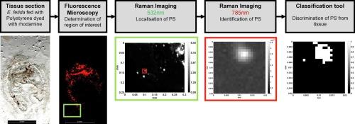
The uptake of microplastic particles (MPP) by organisms is frequently described and poses a potential risk for these organisms and ultimately for humans either through direct uptake or trophic transfer. Currently, the in-situ detection of MPP in organisms is typically based on histological examination of tissue sections after uptake of fluorescently-labelled MPP and is thus not feasible for environmental samples. The alternative approach is purification of MPP from whole organisms or organs by chemical digestion and subsequent spectroscopic detection (FT-IR or Raman). While this approach is feasible for un-labelled particles it goes along with loss of any spatial information related to the location in the tissue. In our study we aimed at providing a workflow for the localisation and identification of non-fluorescent and fluorescent polystyrene (PS) particles (fragments, size range 2–130 μm) in tissue sections of the model organism Eisenia fetida with Raman spectroscopic imaging (RSI). We provide methodological approaches for the preparation of the samples, technical parameters for the RSI measurements and data analysis for PS differentiation in tissue sections. The developed approaches were combined in a workflow for the in-situ analysis of MPP in tissue sections. The spectroscopic analysis requires differentiation of spectra of MPP and interfering compounds, which is challenging given the complexity of tissue. Therefore, a classification algorithm was developed to differentiate PS particles from haem, intestinal contents and surrounding tissue. It allows the differentiation of PS particles from protein in the tissue of E. fetida with an accuracy of 95%. The smallest PS particle detected in the tissue was 2 μm in diameter. We show that it is possible to localise and identify non-fluorescent and fluorescent ingested PS particles directly in tissue sections of E. fetida in the gut lumen and the adjacent tissue.
期刊介绍:
NanoImpact is a multidisciplinary journal that focuses on nanosafety research and areas related to the impacts of manufactured nanomaterials on human and environmental systems and the behavior of nanomaterials in these systems.
 求助内容:
求助内容: 应助结果提醒方式:


