{"title":"严重受损肝细胞启动子区的去甲基化增强趋化因子受体CXCR4基因的表达。","authors":"Chihiro Ito, Ryuma Haraguchi, Kohei Ogawa, Miku Iwata, Riko Kitazawa, Yasutsugu Takada, Sohei Kitazawa","doi":"10.1007/s00418-023-02229-x","DOIUrl":null,"url":null,"abstract":"<p><p>The liver is known to possess remarkable regenerative potential, but persistent inflammation or severe acute injury can lead to liver fibrosis and incomplete regeneration, ultimately resulting in liver failure. Recent studies have shown that the axis of two types of CXCL12 receptors, CXCR4 and CXCR7, plays a crucial role in liver fibrosis and regeneration. The present study aimed to investigate the regulatory factors involved in CXCR4 expression in injured liver. Immunohistochemical screening of liver tissue samples collected during liver transplantation revealed a reciprocal expression pattern between CXCR4 and MeCP2. An in vitro system involving cultured cell lines and H<sub>2</sub>O<sub>2</sub> treatment was established to study the impact of oxidative stress on signaling pathways and epigenetic alterations that affect CXCR4 mRNA expression. Operating through distinct signaling pathways, H<sub>2</sub>O<sub>2</sub> treatment induced a dose-dependent increase in CXCR4 expression in both hepatocyte- and intrahepatic cholangiocyte-derived cells. Treatment of the cells with trichostatin and azacytidine modulated CXCR4 expression in hepatocytes by modifying the methylation status of CpG dinucleotides located in a pair of TA repeats adjacent to the TATA box of the CXCR4 gene promoter. Only MeCP2 bound to oligonucleotides representing the TATA box region when the cytosine residues within the sequence were methylated, as revealed by electrophoretic mobility shift assay (EMSA). Methylation-specific PCR analysis of microdissected samples revealed a correlation between the loss of CpG methylation and the upregulation of CXCR4 in injured hepatocytes, replicating the findings from the in vitro study. Besides the conventional MEK/ERK and NF-κB signaling pathways that activate CXCR4 in intrahepatic cholangiocytes, the unique epigenetic modifications observed in hepatocytes might also contribute to a shift in the CXCR4-CXCR7 balance towards CXCR4, leading to irreversible liver injury and fibrosis. This study highlights the importance of epigenetic modifications in regulating CXCR4 expression in liver injury and fibrosis.</p>","PeriodicalId":13107,"journal":{"name":"Histochemistry and Cell Biology","volume":null,"pages":null},"PeriodicalIF":2.1000,"publicationDate":"2023-11-01","publicationTypes":"Journal Article","fieldsOfStudy":null,"isOpenAccess":false,"openAccessPdf":"","citationCount":"0","resultStr":"{\"title\":\"Demethylation in promoter region of severely damaged hepatocytes enhances chemokine receptor CXCR4 gene expression.\",\"authors\":\"Chihiro Ito, Ryuma Haraguchi, Kohei Ogawa, Miku Iwata, Riko Kitazawa, Yasutsugu Takada, Sohei Kitazawa\",\"doi\":\"10.1007/s00418-023-02229-x\",\"DOIUrl\":null,\"url\":null,\"abstract\":\"<p><p>The liver is known to possess remarkable regenerative potential, but persistent inflammation or severe acute injury can lead to liver fibrosis and incomplete regeneration, ultimately resulting in liver failure. Recent studies have shown that the axis of two types of CXCL12 receptors, CXCR4 and CXCR7, plays a crucial role in liver fibrosis and regeneration. The present study aimed to investigate the regulatory factors involved in CXCR4 expression in injured liver. Immunohistochemical screening of liver tissue samples collected during liver transplantation revealed a reciprocal expression pattern between CXCR4 and MeCP2. An in vitro system involving cultured cell lines and H<sub>2</sub>O<sub>2</sub> treatment was established to study the impact of oxidative stress on signaling pathways and epigenetic alterations that affect CXCR4 mRNA expression. Operating through distinct signaling pathways, H<sub>2</sub>O<sub>2</sub> treatment induced a dose-dependent increase in CXCR4 expression in both hepatocyte- and intrahepatic cholangiocyte-derived cells. Treatment of the cells with trichostatin and azacytidine modulated CXCR4 expression in hepatocytes by modifying the methylation status of CpG dinucleotides located in a pair of TA repeats adjacent to the TATA box of the CXCR4 gene promoter. Only MeCP2 bound to oligonucleotides representing the TATA box region when the cytosine residues within the sequence were methylated, as revealed by electrophoretic mobility shift assay (EMSA). Methylation-specific PCR analysis of microdissected samples revealed a correlation between the loss of CpG methylation and the upregulation of CXCR4 in injured hepatocytes, replicating the findings from the in vitro study. Besides the conventional MEK/ERK and NF-κB signaling pathways that activate CXCR4 in intrahepatic cholangiocytes, the unique epigenetic modifications observed in hepatocytes might also contribute to a shift in the CXCR4-CXCR7 balance towards CXCR4, leading to irreversible liver injury and fibrosis. This study highlights the importance of epigenetic modifications in regulating CXCR4 expression in liver injury and fibrosis.</p>\",\"PeriodicalId\":13107,\"journal\":{\"name\":\"Histochemistry and Cell Biology\",\"volume\":null,\"pages\":null},\"PeriodicalIF\":2.1000,\"publicationDate\":\"2023-11-01\",\"publicationTypes\":\"Journal Article\",\"fieldsOfStudy\":null,\"isOpenAccess\":false,\"openAccessPdf\":\"\",\"citationCount\":\"0\",\"resultStr\":null,\"platform\":\"Semanticscholar\",\"paperid\":null,\"PeriodicalName\":\"Histochemistry and Cell Biology\",\"FirstCategoryId\":\"99\",\"ListUrlMain\":\"https://doi.org/10.1007/s00418-023-02229-x\",\"RegionNum\":4,\"RegionCategory\":\"生物学\",\"ArticlePicture\":[],\"TitleCN\":null,\"AbstractTextCN\":null,\"PMCID\":null,\"EPubDate\":\"2023/8/2 0:00:00\",\"PubModel\":\"Epub\",\"JCR\":\"Q4\",\"JCRName\":\"CELL BIOLOGY\",\"Score\":null,\"Total\":0}","platform":"Semanticscholar","paperid":null,"PeriodicalName":"Histochemistry and Cell Biology","FirstCategoryId":"99","ListUrlMain":"https://doi.org/10.1007/s00418-023-02229-x","RegionNum":4,"RegionCategory":"生物学","ArticlePicture":[],"TitleCN":null,"AbstractTextCN":null,"PMCID":null,"EPubDate":"2023/8/2 0:00:00","PubModel":"Epub","JCR":"Q4","JCRName":"CELL BIOLOGY","Score":null,"Total":0}
Demethylation in promoter region of severely damaged hepatocytes enhances chemokine receptor CXCR4 gene expression.
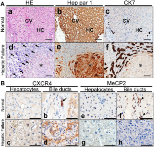
The liver is known to possess remarkable regenerative potential, but persistent inflammation or severe acute injury can lead to liver fibrosis and incomplete regeneration, ultimately resulting in liver failure. Recent studies have shown that the axis of two types of CXCL12 receptors, CXCR4 and CXCR7, plays a crucial role in liver fibrosis and regeneration. The present study aimed to investigate the regulatory factors involved in CXCR4 expression in injured liver. Immunohistochemical screening of liver tissue samples collected during liver transplantation revealed a reciprocal expression pattern between CXCR4 and MeCP2. An in vitro system involving cultured cell lines and H2O2 treatment was established to study the impact of oxidative stress on signaling pathways and epigenetic alterations that affect CXCR4 mRNA expression. Operating through distinct signaling pathways, H2O2 treatment induced a dose-dependent increase in CXCR4 expression in both hepatocyte- and intrahepatic cholangiocyte-derived cells. Treatment of the cells with trichostatin and azacytidine modulated CXCR4 expression in hepatocytes by modifying the methylation status of CpG dinucleotides located in a pair of TA repeats adjacent to the TATA box of the CXCR4 gene promoter. Only MeCP2 bound to oligonucleotides representing the TATA box region when the cytosine residues within the sequence were methylated, as revealed by electrophoretic mobility shift assay (EMSA). Methylation-specific PCR analysis of microdissected samples revealed a correlation between the loss of CpG methylation and the upregulation of CXCR4 in injured hepatocytes, replicating the findings from the in vitro study. Besides the conventional MEK/ERK and NF-κB signaling pathways that activate CXCR4 in intrahepatic cholangiocytes, the unique epigenetic modifications observed in hepatocytes might also contribute to a shift in the CXCR4-CXCR7 balance towards CXCR4, leading to irreversible liver injury and fibrosis. This study highlights the importance of epigenetic modifications in regulating CXCR4 expression in liver injury and fibrosis.
期刊介绍:
Histochemistry and Cell Biology is devoted to the field of molecular histology and cell biology, publishing original articles dealing with the localization and identification of molecular components, metabolic activities and cell biological aspects of cells and tissues. Coverage extends to the development, application, and/or evaluation of methods and probes that can be used in the entire area of histochemistry and cell biology.
 求助内容:
求助内容: 应助结果提醒方式:


