Fatih Ates, Mesut Sivri, Mehmet Sedat Durmaz, Tamer Sekmenli, Metin Gunduz, Ilhan Ciftci
{"title":"传统多普勒成像技术与高超微血管成像技术测定隐睾血管化的比较。","authors":"Fatih Ates, Mesut Sivri, Mehmet Sedat Durmaz, Tamer Sekmenli, Metin Gunduz, Ilhan Ciftci","doi":"10.15557/jou.2023.0013","DOIUrl":null,"url":null,"abstract":"<p><strong>Aim: </strong>Our aim was to gain an idea about testicular injury by comparing the reduced volume, which is one of the indirect indicators of testicular damage in undescended testes, and by evaluating the reduced microvascular blood flow by superb microvascular imaging, and also to determine whether superb microvascular imaging modes could detect microvascular blood flow in more detail in the decreased volume of undescended testes.</p><p><strong>Material and methods: </strong>We compared testicular blood flow in undescended testes via conventional Doppler imaging, color superb microvascular imaging, and monochrome superb microvascular imaging techniques with contralateral normally located testis and normal control group. Each sample of testicular tissue was evaluated using a qualitative method. Spot color encoding and linear flow color encoding counts determined in testicular parenchyma were counted separately and expressed as numerical data. The localization of the examined testes in the grayscale was noted (proximal inguinal canal, medial inguinal canal, distal inguinal canal, and scrotal). The volume of undescended testes was calculated automatically via a formula for volume.</p><p><strong>Results: </strong>Monochrome superb microvascular imaging is significantly superior in visualizing the vascularity of undescended testes compared with color Doppler, power Doppler and color superb microvascular imaging (<i>p</i> = 0.001). Also, undescended testes have a significantly lower blood flow compared with contralateral normal testes (<i>p</i> = 0.001). The volume of undescended testes was significantly lower than the contralateral normal testes.</p><p><strong>Conclusions: </strong>The volume, structure and blood flow are indirect signs of testicular damage in undescended testes. Monochrome superb microvascular imaging can detect vascularity in undescended testes better than the conventional Doppler imaging technique and color superb microvascular imaging. Based on our findings, we can report that monochrome superb microvascular imaging can be used to evaluate testicular injury and vascularity of undescended testes.</p>","PeriodicalId":45612,"journal":{"name":"Journal of Ultrasonography","volume":"23 93","pages":"e66-e72"},"PeriodicalIF":1.5000,"publicationDate":"2023-06-01","publicationTypes":"Journal Article","fieldsOfStudy":null,"isOpenAccess":false,"openAccessPdf":"https://ftp.ncbi.nlm.nih.gov/pub/pmc/oa_pdf/f9/e8/jou-23-93-jou.2023.0013.PMC10379854.pdf","citationCount":"0","resultStr":"{\"title\":\"Comparison of conventional Doppler imaging techniques and superb microvascular imaging in determination of vascularization in undescended testes.\",\"authors\":\"Fatih Ates, Mesut Sivri, Mehmet Sedat Durmaz, Tamer Sekmenli, Metin Gunduz, Ilhan Ciftci\",\"doi\":\"10.15557/jou.2023.0013\",\"DOIUrl\":null,\"url\":null,\"abstract\":\"<p><strong>Aim: </strong>Our aim was to gain an idea about testicular injury by comparing the reduced volume, which is one of the indirect indicators of testicular damage in undescended testes, and by evaluating the reduced microvascular blood flow by superb microvascular imaging, and also to determine whether superb microvascular imaging modes could detect microvascular blood flow in more detail in the decreased volume of undescended testes.</p><p><strong>Material and methods: </strong>We compared testicular blood flow in undescended testes via conventional Doppler imaging, color superb microvascular imaging, and monochrome superb microvascular imaging techniques with contralateral normally located testis and normal control group. Each sample of testicular tissue was evaluated using a qualitative method. Spot color encoding and linear flow color encoding counts determined in testicular parenchyma were counted separately and expressed as numerical data. The localization of the examined testes in the grayscale was noted (proximal inguinal canal, medial inguinal canal, distal inguinal canal, and scrotal). The volume of undescended testes was calculated automatically via a formula for volume.</p><p><strong>Results: </strong>Monochrome superb microvascular imaging is significantly superior in visualizing the vascularity of undescended testes compared with color Doppler, power Doppler and color superb microvascular imaging (<i>p</i> = 0.001). Also, undescended testes have a significantly lower blood flow compared with contralateral normal testes (<i>p</i> = 0.001). The volume of undescended testes was significantly lower than the contralateral normal testes.</p><p><strong>Conclusions: </strong>The volume, structure and blood flow are indirect signs of testicular damage in undescended testes. Monochrome superb microvascular imaging can detect vascularity in undescended testes better than the conventional Doppler imaging technique and color superb microvascular imaging. Based on our findings, we can report that monochrome superb microvascular imaging can be used to evaluate testicular injury and vascularity of undescended testes.</p>\",\"PeriodicalId\":45612,\"journal\":{\"name\":\"Journal of Ultrasonography\",\"volume\":\"23 93\",\"pages\":\"e66-e72\"},\"PeriodicalIF\":1.5000,\"publicationDate\":\"2023-06-01\",\"publicationTypes\":\"Journal Article\",\"fieldsOfStudy\":null,\"isOpenAccess\":false,\"openAccessPdf\":\"https://ftp.ncbi.nlm.nih.gov/pub/pmc/oa_pdf/f9/e8/jou-23-93-jou.2023.0013.PMC10379854.pdf\",\"citationCount\":\"0\",\"resultStr\":null,\"platform\":\"Semanticscholar\",\"paperid\":null,\"PeriodicalName\":\"Journal of Ultrasonography\",\"FirstCategoryId\":\"1085\",\"ListUrlMain\":\"https://doi.org/10.15557/jou.2023.0013\",\"RegionNum\":0,\"RegionCategory\":null,\"ArticlePicture\":[],\"TitleCN\":null,\"AbstractTextCN\":null,\"PMCID\":null,\"EPubDate\":\"\",\"PubModel\":\"\",\"JCR\":\"Q3\",\"JCRName\":\"RADIOLOGY, NUCLEAR MEDICINE & MEDICAL IMAGING\",\"Score\":null,\"Total\":0}","platform":"Semanticscholar","paperid":null,"PeriodicalName":"Journal of Ultrasonography","FirstCategoryId":"1085","ListUrlMain":"https://doi.org/10.15557/jou.2023.0013","RegionNum":0,"RegionCategory":null,"ArticlePicture":[],"TitleCN":null,"AbstractTextCN":null,"PMCID":null,"EPubDate":"","PubModel":"","JCR":"Q3","JCRName":"RADIOLOGY, NUCLEAR MEDICINE & MEDICAL IMAGING","Score":null,"Total":0}
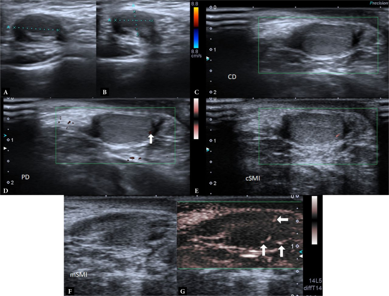
Comparison of conventional Doppler imaging techniques and superb microvascular imaging in determination of vascularization in undescended testes.
Aim: Our aim was to gain an idea about testicular injury by comparing the reduced volume, which is one of the indirect indicators of testicular damage in undescended testes, and by evaluating the reduced microvascular blood flow by superb microvascular imaging, and also to determine whether superb microvascular imaging modes could detect microvascular blood flow in more detail in the decreased volume of undescended testes.
Material and methods: We compared testicular blood flow in undescended testes via conventional Doppler imaging, color superb microvascular imaging, and monochrome superb microvascular imaging techniques with contralateral normally located testis and normal control group. Each sample of testicular tissue was evaluated using a qualitative method. Spot color encoding and linear flow color encoding counts determined in testicular parenchyma were counted separately and expressed as numerical data. The localization of the examined testes in the grayscale was noted (proximal inguinal canal, medial inguinal canal, distal inguinal canal, and scrotal). The volume of undescended testes was calculated automatically via a formula for volume.
Results: Monochrome superb microvascular imaging is significantly superior in visualizing the vascularity of undescended testes compared with color Doppler, power Doppler and color superb microvascular imaging (p = 0.001). Also, undescended testes have a significantly lower blood flow compared with contralateral normal testes (p = 0.001). The volume of undescended testes was significantly lower than the contralateral normal testes.
Conclusions: The volume, structure and blood flow are indirect signs of testicular damage in undescended testes. Monochrome superb microvascular imaging can detect vascularity in undescended testes better than the conventional Doppler imaging technique and color superb microvascular imaging. Based on our findings, we can report that monochrome superb microvascular imaging can be used to evaluate testicular injury and vascularity of undescended testes.

 求助内容:
求助内容: 应助结果提醒方式:


