Marco Migliorati, Anna De Mari, Fabio Annarumma, Hussein Aghazada, Giovanni Battista, Alessandra Campobasso, Maria Menini, Antonino Lo Giudice, Lucia H S Cevidanes, Sara Drago
{"title":"青少年晚期患者骨源性扩张过程中微螺钉位置变化的三维分析。","authors":"Marco Migliorati, Anna De Mari, Fabio Annarumma, Hussein Aghazada, Giovanni Battista, Alessandra Campobasso, Maria Menini, Antonino Lo Giudice, Lucia H S Cevidanes, Sara Drago","doi":"10.1186/s40510-023-00469-0","DOIUrl":null,"url":null,"abstract":"<p><strong>Introduction: </strong>Maxillary expansion in patients at the end of their growth relies on the possibility to use miniscrew supported expanders to apply expansion forces directly to the midpalatal suture. Although miniscrews provide a stable anchorage unit, several studies have reported that they do not remain in exactly the same position during treatment. The aim of the present study was to analyze miniscrew position changes after the expansion using bone-borne appliances in late adolescent patients.</p><p><strong>Methods: </strong>Nineteen patients (13 females, 6 males), with a mean age of 17.81 (SD = 4.66), were treated with a Bone-Borne Expander Device. The appliance was designed with 4 miniscrews: 2 in the anterior palatal area, at the third rugae level; 2 in the posterior area. A CBCT and an intraoral scan were obtained before treatment (T0), and then, a second CBCT was obtained after the expansion (T1). Data on peri-suture bone thickness were collected at T0, then the CBCTs were superimposed, and changes between mini-screws position on T0 and T1 were evaluated, both by linear and angular displacements.</p><p><strong>Results: </strong>Significant longitudinal differences were found in the distance of the head and the tip of miniscrews measured at the occlusal plane, as well as angular changes. Correlations between displacement measurements and peri-suture bone thickness and height measurements were found as well.</p><p><strong>Conclusions: </strong>While acting as bone anchor units, miniscrews do not remain in the same position during bone-borne expansion. The amount of displacement was related to peri-sutural total bone height and cortical thickness, especially in the anterior area of the naso-frontal maxillary complex.</p>","PeriodicalId":56071,"journal":{"name":"Progress in Orthodontics","volume":"24 1","pages":"20"},"PeriodicalIF":4.8000,"publicationDate":"2023-06-05","publicationTypes":"Journal Article","fieldsOfStudy":null,"isOpenAccess":false,"openAccessPdf":"https://www.ncbi.nlm.nih.gov/pmc/articles/PMC10239743/pdf/","citationCount":"2","resultStr":"{\"title\":\"Three-dimensional analysis of miniscrew position changes during bone-borne expansion in young and late adolescent patients.\",\"authors\":\"Marco Migliorati, Anna De Mari, Fabio Annarumma, Hussein Aghazada, Giovanni Battista, Alessandra Campobasso, Maria Menini, Antonino Lo Giudice, Lucia H S Cevidanes, Sara Drago\",\"doi\":\"10.1186/s40510-023-00469-0\",\"DOIUrl\":null,\"url\":null,\"abstract\":\"<p><strong>Introduction: </strong>Maxillary expansion in patients at the end of their growth relies on the possibility to use miniscrew supported expanders to apply expansion forces directly to the midpalatal suture. Although miniscrews provide a stable anchorage unit, several studies have reported that they do not remain in exactly the same position during treatment. The aim of the present study was to analyze miniscrew position changes after the expansion using bone-borne appliances in late adolescent patients.</p><p><strong>Methods: </strong>Nineteen patients (13 females, 6 males), with a mean age of 17.81 (SD = 4.66), were treated with a Bone-Borne Expander Device. The appliance was designed with 4 miniscrews: 2 in the anterior palatal area, at the third rugae level; 2 in the posterior area. A CBCT and an intraoral scan were obtained before treatment (T0), and then, a second CBCT was obtained after the expansion (T1). Data on peri-suture bone thickness were collected at T0, then the CBCTs were superimposed, and changes between mini-screws position on T0 and T1 were evaluated, both by linear and angular displacements.</p><p><strong>Results: </strong>Significant longitudinal differences were found in the distance of the head and the tip of miniscrews measured at the occlusal plane, as well as angular changes. Correlations between displacement measurements and peri-suture bone thickness and height measurements were found as well.</p><p><strong>Conclusions: </strong>While acting as bone anchor units, miniscrews do not remain in the same position during bone-borne expansion. The amount of displacement was related to peri-sutural total bone height and cortical thickness, especially in the anterior area of the naso-frontal maxillary complex.</p>\",\"PeriodicalId\":56071,\"journal\":{\"name\":\"Progress in Orthodontics\",\"volume\":\"24 1\",\"pages\":\"20\"},\"PeriodicalIF\":4.8000,\"publicationDate\":\"2023-06-05\",\"publicationTypes\":\"Journal Article\",\"fieldsOfStudy\":null,\"isOpenAccess\":false,\"openAccessPdf\":\"https://www.ncbi.nlm.nih.gov/pmc/articles/PMC10239743/pdf/\",\"citationCount\":\"2\",\"resultStr\":null,\"platform\":\"Semanticscholar\",\"paperid\":null,\"PeriodicalName\":\"Progress in Orthodontics\",\"FirstCategoryId\":\"3\",\"ListUrlMain\":\"https://doi.org/10.1186/s40510-023-00469-0\",\"RegionNum\":2,\"RegionCategory\":\"医学\",\"ArticlePicture\":[],\"TitleCN\":null,\"AbstractTextCN\":null,\"PMCID\":null,\"EPubDate\":\"\",\"PubModel\":\"\",\"JCR\":\"Q1\",\"JCRName\":\"Dentistry\",\"Score\":null,\"Total\":0}","platform":"Semanticscholar","paperid":null,"PeriodicalName":"Progress in Orthodontics","FirstCategoryId":"3","ListUrlMain":"https://doi.org/10.1186/s40510-023-00469-0","RegionNum":2,"RegionCategory":"医学","ArticlePicture":[],"TitleCN":null,"AbstractTextCN":null,"PMCID":null,"EPubDate":"","PubModel":"","JCR":"Q1","JCRName":"Dentistry","Score":null,"Total":0}
Three-dimensional analysis of miniscrew position changes during bone-borne expansion in young and late adolescent patients.
Introduction: Maxillary expansion in patients at the end of their growth relies on the possibility to use miniscrew supported expanders to apply expansion forces directly to the midpalatal suture. Although miniscrews provide a stable anchorage unit, several studies have reported that they do not remain in exactly the same position during treatment. The aim of the present study was to analyze miniscrew position changes after the expansion using bone-borne appliances in late adolescent patients.
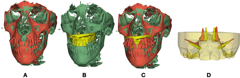
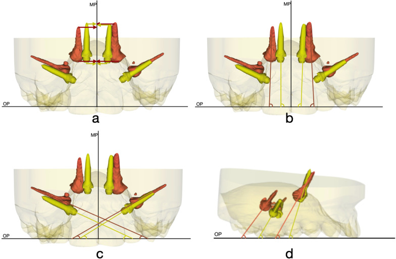
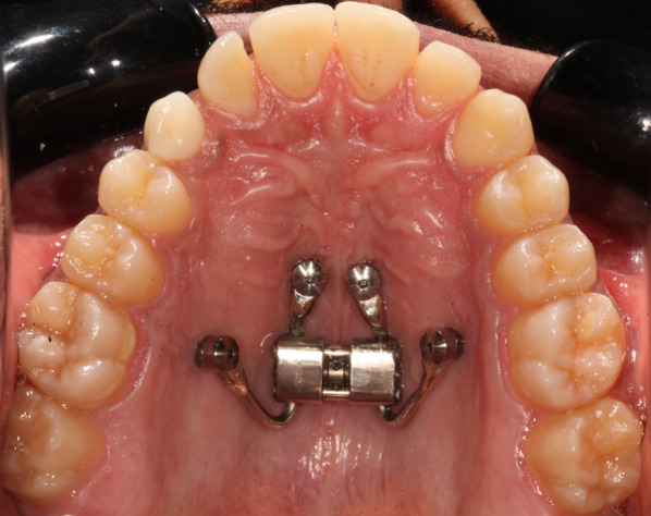
Methods: Nineteen patients (13 females, 6 males), with a mean age of 17.81 (SD = 4.66), were treated with a Bone-Borne Expander Device. The appliance was designed with 4 miniscrews: 2 in the anterior palatal area, at the third rugae level; 2 in the posterior area. A CBCT and an intraoral scan were obtained before treatment (T0), and then, a second CBCT was obtained after the expansion (T1). Data on peri-suture bone thickness were collected at T0, then the CBCTs were superimposed, and changes between mini-screws position on T0 and T1 were evaluated, both by linear and angular displacements.
Results: Significant longitudinal differences were found in the distance of the head and the tip of miniscrews measured at the occlusal plane, as well as angular changes. Correlations between displacement measurements and peri-suture bone thickness and height measurements were found as well.
Conclusions: While acting as bone anchor units, miniscrews do not remain in the same position during bone-borne expansion. The amount of displacement was related to peri-sutural total bone height and cortical thickness, especially in the anterior area of the naso-frontal maxillary complex.
期刊介绍:
Progress in Orthodontics is a fully open access, international journal owned by the Italian Society of Orthodontics and published under the brand SpringerOpen. The Society is currently covering all publication costs so there are no article processing charges for authors.
It is a premier journal of international scope that fosters orthodontic research, including both basic research and development of innovative clinical techniques, with an emphasis on the following areas:
• Mechanisms to improve orthodontics
• Clinical studies and control animal studies
• Orthodontics and genetics, genomics
• Temporomandibular joint (TMJ) control clinical trials
• Efficacy of orthodontic appliances and animal models
• Systematic reviews and meta analyses
• Mechanisms to speed orthodontic treatment
Progress in Orthodontics will consider for publication only meritorious and original contributions. These may be:
• Original articles reporting the findings of clinical trials, clinically relevant basic scientific investigations, or novel therapeutic or diagnostic systems
• Review articles on current topics
• Articles on novel techniques and clinical tools
• Articles of contemporary interest
 求助内容:
求助内容: 应助结果提醒方式:


