Işıl Kefeli, Canan Aslı Utine, Mahmut Kaya, Suleyman Kaynak
{"title":"光学相干断层血管造影成像继发于儿童视网膜震颤的中央凹萎缩。","authors":"Işıl Kefeli, Canan Aslı Utine, Mahmut Kaya, Suleyman Kaynak","doi":"10.14744/bej.2023.38233","DOIUrl":null,"url":null,"abstract":"<p><p>Optical coherence tomography-angiography (OCTA) is a fast, reliable, and non-invasive technique for the diagnosis and follow-up of patients with commotio retinae (CR). Severity of the damage to the retinal and choroidal microvasculature in OCTA imaging and the visual prognosis are directly related to the severity of trauma. There are a few published reports on OCTA in CR that shows alterations of the retinal or superficial choroidal vessels and choriocapillary plexus. OCTA imaging seems to be predictive for visual prognosis. Herein, we present a 6-year-old boy, who had blunt trauma to the right eye with a stick during outdoor playing with visual acuity reduction to 0.1 following resolution of the Berlin's edema. In our case, OCTA revealed damage to the outer layers of the retinae and choriocapillaris and resulting in permanent vision loss. OCTA is a non-invasive, rapid, and safe imaging technique that qualitatively and quantitatively analyzes blood flow from the superficial capillary plexus to the choriocapillaris, which can be predictive in the visual prognosis.</p>","PeriodicalId":8740,"journal":{"name":"Beyoglu Eye Journal","volume":"8 2","pages":"128-133"},"PeriodicalIF":0.0000,"publicationDate":"2023-05-01","publicationTypes":"Journal Article","fieldsOfStudy":null,"isOpenAccess":false,"openAccessPdf":"https://ftp.ncbi.nlm.nih.gov/pub/pmc/oa_pdf/f3/dc/BEJ-8-128.PMC10375211.pdf","citationCount":"0","resultStr":"{\"title\":\"Optical Coherence Tomography Angiography Imaging of Foveal Atrophy Secondary to Commotio Retinae in a Pediatric Patient.\",\"authors\":\"Işıl Kefeli, Canan Aslı Utine, Mahmut Kaya, Suleyman Kaynak\",\"doi\":\"10.14744/bej.2023.38233\",\"DOIUrl\":null,\"url\":null,\"abstract\":\"<p><p>Optical coherence tomography-angiography (OCTA) is a fast, reliable, and non-invasive technique for the diagnosis and follow-up of patients with commotio retinae (CR). Severity of the damage to the retinal and choroidal microvasculature in OCTA imaging and the visual prognosis are directly related to the severity of trauma. There are a few published reports on OCTA in CR that shows alterations of the retinal or superficial choroidal vessels and choriocapillary plexus. OCTA imaging seems to be predictive for visual prognosis. Herein, we present a 6-year-old boy, who had blunt trauma to the right eye with a stick during outdoor playing with visual acuity reduction to 0.1 following resolution of the Berlin's edema. In our case, OCTA revealed damage to the outer layers of the retinae and choriocapillaris and resulting in permanent vision loss. OCTA is a non-invasive, rapid, and safe imaging technique that qualitatively and quantitatively analyzes blood flow from the superficial capillary plexus to the choriocapillaris, which can be predictive in the visual prognosis.</p>\",\"PeriodicalId\":8740,\"journal\":{\"name\":\"Beyoglu Eye Journal\",\"volume\":\"8 2\",\"pages\":\"128-133\"},\"PeriodicalIF\":0.0000,\"publicationDate\":\"2023-05-01\",\"publicationTypes\":\"Journal Article\",\"fieldsOfStudy\":null,\"isOpenAccess\":false,\"openAccessPdf\":\"https://ftp.ncbi.nlm.nih.gov/pub/pmc/oa_pdf/f3/dc/BEJ-8-128.PMC10375211.pdf\",\"citationCount\":\"0\",\"resultStr\":null,\"platform\":\"Semanticscholar\",\"paperid\":null,\"PeriodicalName\":\"Beyoglu Eye Journal\",\"FirstCategoryId\":\"1085\",\"ListUrlMain\":\"https://doi.org/10.14744/bej.2023.38233\",\"RegionNum\":0,\"RegionCategory\":null,\"ArticlePicture\":[],\"TitleCN\":null,\"AbstractTextCN\":null,\"PMCID\":null,\"EPubDate\":\"2023/1/1 0:00:00\",\"PubModel\":\"eCollection\",\"JCR\":\"\",\"JCRName\":\"\",\"Score\":null,\"Total\":0}","platform":"Semanticscholar","paperid":null,"PeriodicalName":"Beyoglu Eye Journal","FirstCategoryId":"1085","ListUrlMain":"https://doi.org/10.14744/bej.2023.38233","RegionNum":0,"RegionCategory":null,"ArticlePicture":[],"TitleCN":null,"AbstractTextCN":null,"PMCID":null,"EPubDate":"2023/1/1 0:00:00","PubModel":"eCollection","JCR":"","JCRName":"","Score":null,"Total":0}
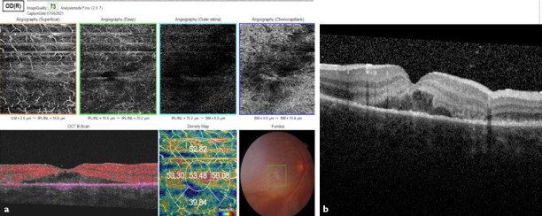
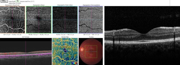
Optical Coherence Tomography Angiography Imaging of Foveal Atrophy Secondary to Commotio Retinae in a Pediatric Patient.
Optical coherence tomography-angiography (OCTA) is a fast, reliable, and non-invasive technique for the diagnosis and follow-up of patients with commotio retinae (CR). Severity of the damage to the retinal and choroidal microvasculature in OCTA imaging and the visual prognosis are directly related to the severity of trauma. There are a few published reports on OCTA in CR that shows alterations of the retinal or superficial choroidal vessels and choriocapillary plexus. OCTA imaging seems to be predictive for visual prognosis. Herein, we present a 6-year-old boy, who had blunt trauma to the right eye with a stick during outdoor playing with visual acuity reduction to 0.1 following resolution of the Berlin's edema. In our case, OCTA revealed damage to the outer layers of the retinae and choriocapillaris and resulting in permanent vision loss. OCTA is a non-invasive, rapid, and safe imaging technique that qualitatively and quantitatively analyzes blood flow from the superficial capillary plexus to the choriocapillaris, which can be predictive in the visual prognosis.


 求助内容:
求助内容: 应助结果提醒方式:


