{"title":"ⅱ类1分错牙合的前磨牙拔除与固定功能治疗的效果及唇廓变化:一项随机对照临床试验。","authors":"Gagan Deep Kochar, Sanjay Londhe, Sukhbir Singh Chopra, Sarvraj Kohli, Virinder Singh Kohli, Ashish Kamboj, Munish Verma","doi":"10.1590/2177-6709.28.2.e232140.oar","DOIUrl":null,"url":null,"abstract":"<p><strong>Objective: </strong>The objective of this two-arm parallel randomized controlled trial was to evaluate the treatment effects and lip profile changes in skeletal Class II patients subjected to premolars extraction treatment versus fixed functional treatment.</p><p><strong>Methods: </strong>Forty six subjects fulfilling inclusion criteria were randomly distributed into Group PE (mean age 13.03±1.78 years) and Group FF (mean age 12.80±1.67 years) (n=23 each). Group PE was managed by therapeutic extraction of maxillary first premolars and mandibular second premolars, followed by mini-implant-supported space closure; and Group FF, by fixed functional appliance therapy. Skeletal, dental, and soft-tissue changes were analyzed using pre and post-treatment lateral cephalograms. Data obtained from this open label study was subjected to blind statistical analysis.</p><p><strong>Results: </strong>Extraction treatment resulted in greater increase of nasolabial angle (NLA: 3.1 [95% CI 2.08, 4.19], p<0.001), significant improvement of upper lip (UL-E line: -2.91 [95% CI -3.54, -2.28], p<0.001, UL-S line: -2.50 [95% CI -2.76, -2.24], p<0.001, UL-SnPog': -2.32 [95% CI -2.90, -1.74], p<0.01) and lower lip position (LL-E line: -0.68 [95% CI -1.36, 0.00], p<0.01, LL-S line: -0.55 [95% CI -1.11, 0.02], p<0.01, and LL-SnPog': -0.64 [95% CI -1.20, -0.07], p<0.01), lip thickness (UL thickness: 2.27 [95% CI 1.79, 2.75], p<0.001; LL thickness: 0.41 [95% CI -0.16, 0.97], p<0.01), upper lip strain (UL strain: -2.68 [95% CI -3.32, -2.04], p<0.001) and soft tissue profile (N'-Sn-Pog': 2.68 [95% CI 1.87, 3.50], p<0.01). No significant difference was observed between the groups regarding skeletal changes in the maxilla and mandible, growth pattern, overjet, overbite, interincisal angle and soft tissue chin position (p>0.05). Premolar extraction treatment demonstrated significant intrusion-retraction of maxillary incisors, better maintenance of maxillary incisor inclination, and significant mandibular molar protraction; whereas functional treatment resulted in retrusive and intrusive effect on maxillary molars, marked proclination of mandibular anterior teeth, and significant extrusion of mandibular molars. Both treatment modalities had similar treatment duration. Implant failure was seen in 7.9% of cases, whereas failure of fixed functional appliance was observed in 9.09% of cases.</p><p><strong>Conclusions: </strong>Premolar extraction therapy is a better treatment modality, compared to fixed functional appliance therapy for Class II patients with moderate skeletal discrepancy, increased overjet, protruded maxillary incisors and protruded lips, as it produces better dentoalveolar response and permits greater improvement of the soft tissue profile and lip relationship.</p>","PeriodicalId":38720,"journal":{"name":"Dental Press Journal of Orthodontics","volume":"28 2","pages":"e232140"},"PeriodicalIF":0.0000,"publicationDate":"2023-01-01","publicationTypes":"Journal Article","fieldsOfStudy":null,"isOpenAccess":false,"openAccessPdf":"https://www.ncbi.nlm.nih.gov/pmc/articles/PMC10202449/pdf/","citationCount":"0","resultStr":"{\"title\":\"Treatment effects and lip profile changes following premolars extraction treatment vs fixed functional treatment in Class II division 1 malocclusion: A randomized controlled clinical trial.\",\"authors\":\"Gagan Deep Kochar, Sanjay Londhe, Sukhbir Singh Chopra, Sarvraj Kohli, Virinder Singh Kohli, Ashish Kamboj, Munish Verma\",\"doi\":\"10.1590/2177-6709.28.2.e232140.oar\",\"DOIUrl\":null,\"url\":null,\"abstract\":\"<p><strong>Objective: </strong>The objective of this two-arm parallel randomized controlled trial was to evaluate the treatment effects and lip profile changes in skeletal Class II patients subjected to premolars extraction treatment versus fixed functional treatment.</p><p><strong>Methods: </strong>Forty six subjects fulfilling inclusion criteria were randomly distributed into Group PE (mean age 13.03±1.78 years) and Group FF (mean age 12.80±1.67 years) (n=23 each). Group PE was managed by therapeutic extraction of maxillary first premolars and mandibular second premolars, followed by mini-implant-supported space closure; and Group FF, by fixed functional appliance therapy. Skeletal, dental, and soft-tissue changes were analyzed using pre and post-treatment lateral cephalograms. Data obtained from this open label study was subjected to blind statistical analysis.</p><p><strong>Results: </strong>Extraction treatment resulted in greater increase of nasolabial angle (NLA: 3.1 [95% CI 2.08, 4.19], p<0.001), significant improvement of upper lip (UL-E line: -2.91 [95% CI -3.54, -2.28], p<0.001, UL-S line: -2.50 [95% CI -2.76, -2.24], p<0.001, UL-SnPog': -2.32 [95% CI -2.90, -1.74], p<0.01) and lower lip position (LL-E line: -0.68 [95% CI -1.36, 0.00], p<0.01, LL-S line: -0.55 [95% CI -1.11, 0.02], p<0.01, and LL-SnPog': -0.64 [95% CI -1.20, -0.07], p<0.01), lip thickness (UL thickness: 2.27 [95% CI 1.79, 2.75], p<0.001; LL thickness: 0.41 [95% CI -0.16, 0.97], p<0.01), upper lip strain (UL strain: -2.68 [95% CI -3.32, -2.04], p<0.001) and soft tissue profile (N'-Sn-Pog': 2.68 [95% CI 1.87, 3.50], p<0.01). No significant difference was observed between the groups regarding skeletal changes in the maxilla and mandible, growth pattern, overjet, overbite, interincisal angle and soft tissue chin position (p>0.05). Premolar extraction treatment demonstrated significant intrusion-retraction of maxillary incisors, better maintenance of maxillary incisor inclination, and significant mandibular molar protraction; whereas functional treatment resulted in retrusive and intrusive effect on maxillary molars, marked proclination of mandibular anterior teeth, and significant extrusion of mandibular molars. Both treatment modalities had similar treatment duration. Implant failure was seen in 7.9% of cases, whereas failure of fixed functional appliance was observed in 9.09% of cases.</p><p><strong>Conclusions: </strong>Premolar extraction therapy is a better treatment modality, compared to fixed functional appliance therapy for Class II patients with moderate skeletal discrepancy, increased overjet, protruded maxillary incisors and protruded lips, as it produces better dentoalveolar response and permits greater improvement of the soft tissue profile and lip relationship.</p>\",\"PeriodicalId\":38720,\"journal\":{\"name\":\"Dental Press Journal of Orthodontics\",\"volume\":\"28 2\",\"pages\":\"e232140\"},\"PeriodicalIF\":0.0000,\"publicationDate\":\"2023-01-01\",\"publicationTypes\":\"Journal Article\",\"fieldsOfStudy\":null,\"isOpenAccess\":false,\"openAccessPdf\":\"https://www.ncbi.nlm.nih.gov/pmc/articles/PMC10202449/pdf/\",\"citationCount\":\"0\",\"resultStr\":null,\"platform\":\"Semanticscholar\",\"paperid\":null,\"PeriodicalName\":\"Dental Press Journal of Orthodontics\",\"FirstCategoryId\":\"1085\",\"ListUrlMain\":\"https://doi.org/10.1590/2177-6709.28.2.e232140.oar\",\"RegionNum\":0,\"RegionCategory\":null,\"ArticlePicture\":[],\"TitleCN\":null,\"AbstractTextCN\":null,\"PMCID\":null,\"EPubDate\":\"\",\"PubModel\":\"\",\"JCR\":\"Q2\",\"JCRName\":\"Medicine\",\"Score\":null,\"Total\":0}","platform":"Semanticscholar","paperid":null,"PeriodicalName":"Dental Press Journal of Orthodontics","FirstCategoryId":"1085","ListUrlMain":"https://doi.org/10.1590/2177-6709.28.2.e232140.oar","RegionNum":0,"RegionCategory":null,"ArticlePicture":[],"TitleCN":null,"AbstractTextCN":null,"PMCID":null,"EPubDate":"","PubModel":"","JCR":"Q2","JCRName":"Medicine","Score":null,"Total":0}
引用次数: 0
摘要
目的:本研究是一项双臂平行随机对照试验,目的是评估骨骼II类患者接受前磨牙拔除治疗与固定功能治疗的治疗效果和唇廓变化。方法:符合入选标准的46例受试者随机分为PE组(平均年龄13.03±1.78岁)和FF组(平均年龄12.80±1.67岁),每组23例。PE组采用治疗性拔除上颌第一前磨牙和下颌第二前磨牙,然后进行微型种植体支撑空隙封闭;FF组采用固定式功能矫治器治疗。使用治疗前后侧位脑电图分析骨骼、牙齿和软组织的变化。从这项开放标签研究中获得的数据进行了盲统计分析。结果:拔牙组鼻唇角增大较大(NLA: 3.1 [95% CI 2.08, 4.19], p0.05)。前磨牙拔除后上颌切牙的内缩明显,上颌切牙的倾斜度维持较好,下颌磨牙的前伸明显;而功能性治疗导致上颌磨牙的后移和侵入作用,下颌前牙明显前倾,下颌磨牙明显挤压。两种治疗方式的治疗时间相似。7.9%的病例出现假体失败,而9.09%的病例出现固定功能矫治器失败。结论:对于中等骨骼差异、覆盖增加、上颌门牙突出、嘴唇突出的II类患者,前磨牙拔牙治疗比固定功能矫治治疗效果更好,牙槽反应更好,软组织形态和唇部关系改善更大。
Treatment effects and lip profile changes following premolars extraction treatment vs fixed functional treatment in Class II division 1 malocclusion: A randomized controlled clinical trial.
Objective: The objective of this two-arm parallel randomized controlled trial was to evaluate the treatment effects and lip profile changes in skeletal Class II patients subjected to premolars extraction treatment versus fixed functional treatment.
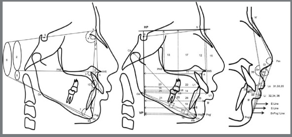
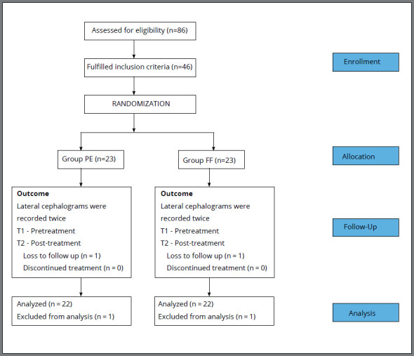
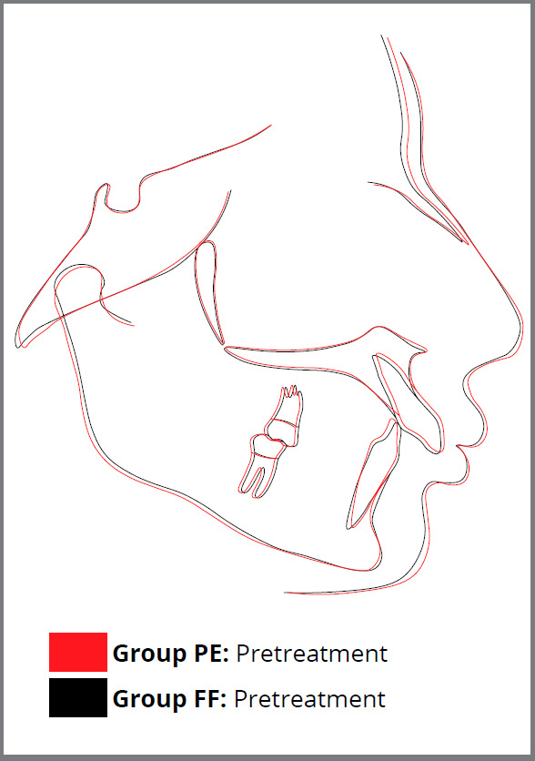
Methods: Forty six subjects fulfilling inclusion criteria were randomly distributed into Group PE (mean age 13.03±1.78 years) and Group FF (mean age 12.80±1.67 years) (n=23 each). Group PE was managed by therapeutic extraction of maxillary first premolars and mandibular second premolars, followed by mini-implant-supported space closure; and Group FF, by fixed functional appliance therapy. Skeletal, dental, and soft-tissue changes were analyzed using pre and post-treatment lateral cephalograms. Data obtained from this open label study was subjected to blind statistical analysis.
Results: Extraction treatment resulted in greater increase of nasolabial angle (NLA: 3.1 [95% CI 2.08, 4.19], p<0.001), significant improvement of upper lip (UL-E line: -2.91 [95% CI -3.54, -2.28], p<0.001, UL-S line: -2.50 [95% CI -2.76, -2.24], p<0.001, UL-SnPog': -2.32 [95% CI -2.90, -1.74], p<0.01) and lower lip position (LL-E line: -0.68 [95% CI -1.36, 0.00], p<0.01, LL-S line: -0.55 [95% CI -1.11, 0.02], p<0.01, and LL-SnPog': -0.64 [95% CI -1.20, -0.07], p<0.01), lip thickness (UL thickness: 2.27 [95% CI 1.79, 2.75], p<0.001; LL thickness: 0.41 [95% CI -0.16, 0.97], p<0.01), upper lip strain (UL strain: -2.68 [95% CI -3.32, -2.04], p<0.001) and soft tissue profile (N'-Sn-Pog': 2.68 [95% CI 1.87, 3.50], p<0.01). No significant difference was observed between the groups regarding skeletal changes in the maxilla and mandible, growth pattern, overjet, overbite, interincisal angle and soft tissue chin position (p>0.05). Premolar extraction treatment demonstrated significant intrusion-retraction of maxillary incisors, better maintenance of maxillary incisor inclination, and significant mandibular molar protraction; whereas functional treatment resulted in retrusive and intrusive effect on maxillary molars, marked proclination of mandibular anterior teeth, and significant extrusion of mandibular molars. Both treatment modalities had similar treatment duration. Implant failure was seen in 7.9% of cases, whereas failure of fixed functional appliance was observed in 9.09% of cases.
Conclusions: Premolar extraction therapy is a better treatment modality, compared to fixed functional appliance therapy for Class II patients with moderate skeletal discrepancy, increased overjet, protruded maxillary incisors and protruded lips, as it produces better dentoalveolar response and permits greater improvement of the soft tissue profile and lip relationship.
期刊介绍:
The Dental Press Journal of Orthodontics publishes scientific research articles, significant reviews, clinical and technical case reports, brief communications, and other materials related to Orthodontics and Facial Orthopedics.

 求助内容:
求助内容: 应助结果提醒方式:


