Claire Benwood, Jonathan Walters-Shumka, Kali Scheck, Stephanie M Willerth
{"title":"使用智能生物链接3D生物打印患者来源的诱导多能干细胞阿尔茨海默病模型。","authors":"Claire Benwood, Jonathan Walters-Shumka, Kali Scheck, Stephanie M Willerth","doi":"10.1186/s42234-023-00112-7","DOIUrl":null,"url":null,"abstract":"<p><strong>Background: </strong>Alzheimer's disease (AD), a progressive neurodegenerative disorder, is becoming increasingly prevalent as our population ages. It is characterized by the buildup of amyloid beta plaques and neurofibrillary tangles containing hyperphosphorylated-tau. The current treatments for AD do not prevent the long-term progression of the disease and pre-clinical models often do not accurately represent its complexity. Bioprinting combines cells and biomaterials to create 3D structures that replicate the native tissue environment and can be used as a tool in disease modeling or drug screening.</p><p><strong>Methods: </strong>This work differentiated both healthy and diseased patient-derived human induced pluripotent stems cells (hiPSCs) into neural progenitor cells (NPCs) that were bioprinted using the Aspect RX1 microfluidic printer into dome-shaped constructs. The combination of cells, bioink, and puromorphamine (puro)-releasing microspheres were used to mimic the in vivo environment and direct the differentiation of the NPCs into basal forebrain-resembling cholinergic neurons (BFCN). These tissue models were then characterized for cell viability, immunocytochemistry, and electrophysiology to evaluate their functionality and physiology for use as disease-specific neural models.</p><p><strong>Results: </strong>Tissue models were successfully bioprinted and the cells were viable for analysis after 30- and 45-day cultures. The neuronal and cholinergic markers β-tubulin III (Tuj1), forkhead box G1 (FOXG1), and choline acetyltransferase (ChAT) were identified as well as the AD markers amyloid beta and tau. Further, immature electrical activity was observed when the cells were excited with potassium chloride and acetylcholine.</p><p><strong>Conclusions: </strong>This work shows the successful development of bioprinted tissue models incorporating patient derived hiPSCs. Such models can potentially be used as a tool to screen promising drug candidates for treating AD. Further, this model could be used to increase the understanding of AD progression. The use of patient derived cells also shows the potential of this model for use in personalized medicine applications.</p>","PeriodicalId":72363,"journal":{"name":"Bioelectronic medicine","volume":"9 1","pages":"10"},"PeriodicalIF":0.0000,"publicationDate":"2023-05-24","publicationTypes":"Journal Article","fieldsOfStudy":null,"isOpenAccess":false,"openAccessPdf":"https://www.ncbi.nlm.nih.gov/pmc/articles/PMC10207712/pdf/","citationCount":"0","resultStr":"{\"title\":\"3D bioprinting patient-derived induced pluripotent stem cell models of Alzheimer's disease using a smart bioink.\",\"authors\":\"Claire Benwood, Jonathan Walters-Shumka, Kali Scheck, Stephanie M Willerth\",\"doi\":\"10.1186/s42234-023-00112-7\",\"DOIUrl\":null,\"url\":null,\"abstract\":\"<p><strong>Background: </strong>Alzheimer's disease (AD), a progressive neurodegenerative disorder, is becoming increasingly prevalent as our population ages. It is characterized by the buildup of amyloid beta plaques and neurofibrillary tangles containing hyperphosphorylated-tau. The current treatments for AD do not prevent the long-term progression of the disease and pre-clinical models often do not accurately represent its complexity. Bioprinting combines cells and biomaterials to create 3D structures that replicate the native tissue environment and can be used as a tool in disease modeling or drug screening.</p><p><strong>Methods: </strong>This work differentiated both healthy and diseased patient-derived human induced pluripotent stems cells (hiPSCs) into neural progenitor cells (NPCs) that were bioprinted using the Aspect RX1 microfluidic printer into dome-shaped constructs. The combination of cells, bioink, and puromorphamine (puro)-releasing microspheres were used to mimic the in vivo environment and direct the differentiation of the NPCs into basal forebrain-resembling cholinergic neurons (BFCN). These tissue models were then characterized for cell viability, immunocytochemistry, and electrophysiology to evaluate their functionality and physiology for use as disease-specific neural models.</p><p><strong>Results: </strong>Tissue models were successfully bioprinted and the cells were viable for analysis after 30- and 45-day cultures. The neuronal and cholinergic markers β-tubulin III (Tuj1), forkhead box G1 (FOXG1), and choline acetyltransferase (ChAT) were identified as well as the AD markers amyloid beta and tau. Further, immature electrical activity was observed when the cells were excited with potassium chloride and acetylcholine.</p><p><strong>Conclusions: </strong>This work shows the successful development of bioprinted tissue models incorporating patient derived hiPSCs. Such models can potentially be used as a tool to screen promising drug candidates for treating AD. Further, this model could be used to increase the understanding of AD progression. The use of patient derived cells also shows the potential of this model for use in personalized medicine applications.</p>\",\"PeriodicalId\":72363,\"journal\":{\"name\":\"Bioelectronic medicine\",\"volume\":\"9 1\",\"pages\":\"10\"},\"PeriodicalIF\":0.0000,\"publicationDate\":\"2023-05-24\",\"publicationTypes\":\"Journal Article\",\"fieldsOfStudy\":null,\"isOpenAccess\":false,\"openAccessPdf\":\"https://www.ncbi.nlm.nih.gov/pmc/articles/PMC10207712/pdf/\",\"citationCount\":\"0\",\"resultStr\":null,\"platform\":\"Semanticscholar\",\"paperid\":null,\"PeriodicalName\":\"Bioelectronic medicine\",\"FirstCategoryId\":\"1085\",\"ListUrlMain\":\"https://doi.org/10.1186/s42234-023-00112-7\",\"RegionNum\":0,\"RegionCategory\":null,\"ArticlePicture\":[],\"TitleCN\":null,\"AbstractTextCN\":null,\"PMCID\":null,\"EPubDate\":\"\",\"PubModel\":\"\",\"JCR\":\"\",\"JCRName\":\"\",\"Score\":null,\"Total\":0}","platform":"Semanticscholar","paperid":null,"PeriodicalName":"Bioelectronic medicine","FirstCategoryId":"1085","ListUrlMain":"https://doi.org/10.1186/s42234-023-00112-7","RegionNum":0,"RegionCategory":null,"ArticlePicture":[],"TitleCN":null,"AbstractTextCN":null,"PMCID":null,"EPubDate":"","PubModel":"","JCR":"","JCRName":"","Score":null,"Total":0}
3D bioprinting patient-derived induced pluripotent stem cell models of Alzheimer's disease using a smart bioink.
Background: Alzheimer's disease (AD), a progressive neurodegenerative disorder, is becoming increasingly prevalent as our population ages. It is characterized by the buildup of amyloid beta plaques and neurofibrillary tangles containing hyperphosphorylated-tau. The current treatments for AD do not prevent the long-term progression of the disease and pre-clinical models often do not accurately represent its complexity. Bioprinting combines cells and biomaterials to create 3D structures that replicate the native tissue environment and can be used as a tool in disease modeling or drug screening.
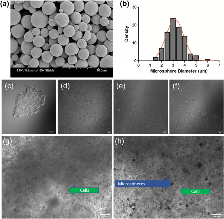
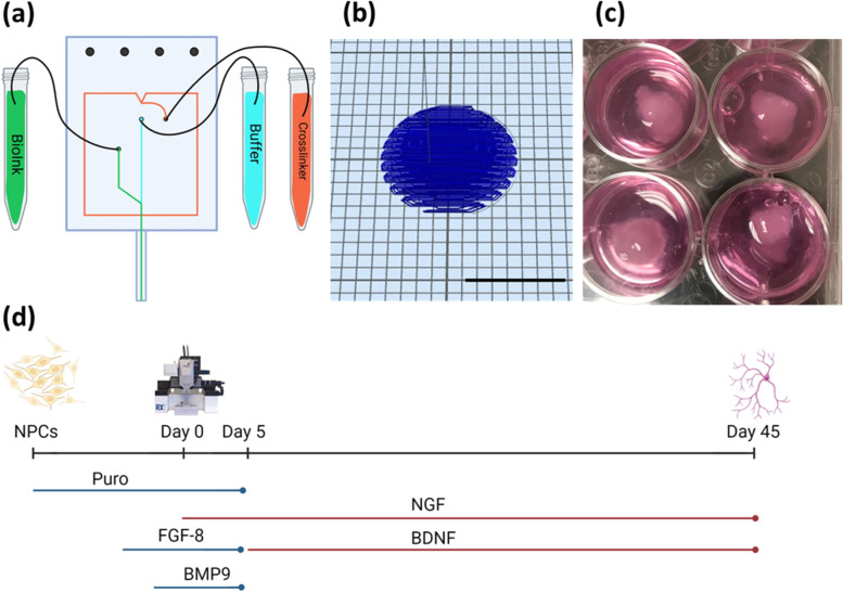
Methods: This work differentiated both healthy and diseased patient-derived human induced pluripotent stems cells (hiPSCs) into neural progenitor cells (NPCs) that were bioprinted using the Aspect RX1 microfluidic printer into dome-shaped constructs. The combination of cells, bioink, and puromorphamine (puro)-releasing microspheres were used to mimic the in vivo environment and direct the differentiation of the NPCs into basal forebrain-resembling cholinergic neurons (BFCN). These tissue models were then characterized for cell viability, immunocytochemistry, and electrophysiology to evaluate their functionality and physiology for use as disease-specific neural models.
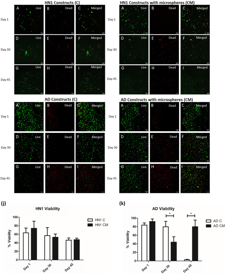
Results: Tissue models were successfully bioprinted and the cells were viable for analysis after 30- and 45-day cultures. The neuronal and cholinergic markers β-tubulin III (Tuj1), forkhead box G1 (FOXG1), and choline acetyltransferase (ChAT) were identified as well as the AD markers amyloid beta and tau. Further, immature electrical activity was observed when the cells were excited with potassium chloride and acetylcholine.
Conclusions: This work shows the successful development of bioprinted tissue models incorporating patient derived hiPSCs. Such models can potentially be used as a tool to screen promising drug candidates for treating AD. Further, this model could be used to increase the understanding of AD progression. The use of patient derived cells also shows the potential of this model for use in personalized medicine applications.

 求助内容:
求助内容: 应助结果提醒方式:


