Yongfeng Li, Huawei Liu, Chao Wang, Rongzeng Yan, Lei Xiang, Xiaodan Mu, Lingling Zheng, Changkui Liu, Min Hu
{"title":"3D打印钛网格支架促进下颌节段性缺损成骨。","authors":"Yongfeng Li, Huawei Liu, Chao Wang, Rongzeng Yan, Lei Xiang, Xiaodan Mu, Lingling Zheng, Changkui Liu, Min Hu","doi":"10.1038/s41536-023-00308-0","DOIUrl":null,"url":null,"abstract":"<p><p>Bone fusion of defect broken ends is the basis of the functional reconstruction of critical maxillofacial segmental bone defects. However, the currently available treatments do not easily achieve this goal. Therefore, this study aimed to fabricate 3D-printing titanium grid scaffolds, which possess sufficient pores and basic biomechanical strength to facilitate osteogenesis in order to accomplish bone fusion in mandibular segmental bone defects. The clinical trial was approved and supervised by the Medical Ethics Committee of the Chinese PLA General Hospital on March 28th, 2019 (Beijing, China. approval No. S2019-065-01), and registered in the clinical trials registry platform (registration number: ChiCTR2300072209). Titanium grid scaffolds were manufactured using selective laser melting and implanted in 20 beagle dogs with mandibular segmental defects. Half of the animals were treated with autologous bone chips and bone substances incorporated into the scaffolds; no additional filling was used for the rest of the animals. After 18 months of observation, radiological scanning and histological analysis in canine models revealed that the pores of regenerated bone were filled with titanium grid scaffolds and bone broken ends were integrated. Furthermore, three patients were treated with similar titanium grid scaffold implants in mandibular segmental defects; no mechanical complications were observed, and similar bone regeneration was observed in the reconstructed patients' mandibles in the clinic. These results demonstrated that 3D-printing titanium grid scaffolds with sufficient pores and basic biomechanical strength could facilitate bone regeneration in large-segment mandibular bone defects.</p>","PeriodicalId":54236,"journal":{"name":"npj Regenerative Medicine","volume":"8 1","pages":"38"},"PeriodicalIF":6.4000,"publicationDate":"2023-07-24","publicationTypes":"Journal Article","fieldsOfStudy":null,"isOpenAccess":false,"openAccessPdf":"https://www.ncbi.nlm.nih.gov/pmc/articles/PMC10366137/pdf/","citationCount":"3","resultStr":"{\"title\":\"3D printing titanium grid scaffold facilitates osteogenesis in mandibular segmental defects.\",\"authors\":\"Yongfeng Li, Huawei Liu, Chao Wang, Rongzeng Yan, Lei Xiang, Xiaodan Mu, Lingling Zheng, Changkui Liu, Min Hu\",\"doi\":\"10.1038/s41536-023-00308-0\",\"DOIUrl\":null,\"url\":null,\"abstract\":\"<p><p>Bone fusion of defect broken ends is the basis of the functional reconstruction of critical maxillofacial segmental bone defects. However, the currently available treatments do not easily achieve this goal. Therefore, this study aimed to fabricate 3D-printing titanium grid scaffolds, which possess sufficient pores and basic biomechanical strength to facilitate osteogenesis in order to accomplish bone fusion in mandibular segmental bone defects. The clinical trial was approved and supervised by the Medical Ethics Committee of the Chinese PLA General Hospital on March 28th, 2019 (Beijing, China. approval No. S2019-065-01), and registered in the clinical trials registry platform (registration number: ChiCTR2300072209). Titanium grid scaffolds were manufactured using selective laser melting and implanted in 20 beagle dogs with mandibular segmental defects. Half of the animals were treated with autologous bone chips and bone substances incorporated into the scaffolds; no additional filling was used for the rest of the animals. After 18 months of observation, radiological scanning and histological analysis in canine models revealed that the pores of regenerated bone were filled with titanium grid scaffolds and bone broken ends were integrated. Furthermore, three patients were treated with similar titanium grid scaffold implants in mandibular segmental defects; no mechanical complications were observed, and similar bone regeneration was observed in the reconstructed patients' mandibles in the clinic. These results demonstrated that 3D-printing titanium grid scaffolds with sufficient pores and basic biomechanical strength could facilitate bone regeneration in large-segment mandibular bone defects.</p>\",\"PeriodicalId\":54236,\"journal\":{\"name\":\"npj Regenerative Medicine\",\"volume\":\"8 1\",\"pages\":\"38\"},\"PeriodicalIF\":6.4000,\"publicationDate\":\"2023-07-24\",\"publicationTypes\":\"Journal Article\",\"fieldsOfStudy\":null,\"isOpenAccess\":false,\"openAccessPdf\":\"https://www.ncbi.nlm.nih.gov/pmc/articles/PMC10366137/pdf/\",\"citationCount\":\"3\",\"resultStr\":null,\"platform\":\"Semanticscholar\",\"paperid\":null,\"PeriodicalName\":\"npj Regenerative Medicine\",\"FirstCategoryId\":\"3\",\"ListUrlMain\":\"https://doi.org/10.1038/s41536-023-00308-0\",\"RegionNum\":1,\"RegionCategory\":\"医学\",\"ArticlePicture\":[],\"TitleCN\":null,\"AbstractTextCN\":null,\"PMCID\":null,\"EPubDate\":\"\",\"PubModel\":\"\",\"JCR\":\"Q1\",\"JCRName\":\"CELL & TISSUE ENGINEERING\",\"Score\":null,\"Total\":0}","platform":"Semanticscholar","paperid":null,"PeriodicalName":"npj Regenerative Medicine","FirstCategoryId":"3","ListUrlMain":"https://doi.org/10.1038/s41536-023-00308-0","RegionNum":1,"RegionCategory":"医学","ArticlePicture":[],"TitleCN":null,"AbstractTextCN":null,"PMCID":null,"EPubDate":"","PubModel":"","JCR":"Q1","JCRName":"CELL & TISSUE ENGINEERING","Score":null,"Total":0}
3D printing titanium grid scaffold facilitates osteogenesis in mandibular segmental defects.
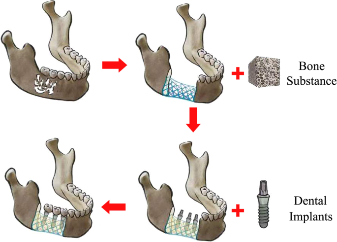
Bone fusion of defect broken ends is the basis of the functional reconstruction of critical maxillofacial segmental bone defects. However, the currently available treatments do not easily achieve this goal. Therefore, this study aimed to fabricate 3D-printing titanium grid scaffolds, which possess sufficient pores and basic biomechanical strength to facilitate osteogenesis in order to accomplish bone fusion in mandibular segmental bone defects. The clinical trial was approved and supervised by the Medical Ethics Committee of the Chinese PLA General Hospital on March 28th, 2019 (Beijing, China. approval No. S2019-065-01), and registered in the clinical trials registry platform (registration number: ChiCTR2300072209). Titanium grid scaffolds were manufactured using selective laser melting and implanted in 20 beagle dogs with mandibular segmental defects. Half of the animals were treated with autologous bone chips and bone substances incorporated into the scaffolds; no additional filling was used for the rest of the animals. After 18 months of observation, radiological scanning and histological analysis in canine models revealed that the pores of regenerated bone were filled with titanium grid scaffolds and bone broken ends were integrated. Furthermore, three patients were treated with similar titanium grid scaffold implants in mandibular segmental defects; no mechanical complications were observed, and similar bone regeneration was observed in the reconstructed patients' mandibles in the clinic. These results demonstrated that 3D-printing titanium grid scaffolds with sufficient pores and basic biomechanical strength could facilitate bone regeneration in large-segment mandibular bone defects.
期刊介绍:
Regenerative Medicine, an innovative online-only journal, aims to advance research in the field of repairing and regenerating damaged tissues and organs within the human body. As a part of the prestigious Nature Partner Journals series and in partnership with ARMI, this high-quality, open access journal serves as a platform for scientists to explore effective therapies that harness the body's natural regenerative capabilities. With a focus on understanding the fundamental mechanisms of tissue damage and regeneration, npj Regenerative Medicine actively encourages studies that bridge the gap between basic research and clinical tissue repair strategies.

 求助内容:
求助内容: 应助结果提醒方式:


