Michal Schäfer, Max B Mitchell, Caitlin Brateng, D Dunbar Ivy, Kendall S Hunter, Dustin B Nash, Johannes C von Alvensleben
{"title":"从标准临床便携式文档格式文件中提取和数字化心电图信号,用于T波形态的主成分分析。","authors":"Michal Schäfer, Max B Mitchell, Caitlin Brateng, D Dunbar Ivy, Kendall S Hunter, Dustin B Nash, Johannes C von Alvensleben","doi":"10.1007/s13239-023-00673-3","DOIUrl":null,"url":null,"abstract":"<p><strong>Introduction: </strong>T-wave analysis from standard electrocardiogram (ECG) remains one of the most available clinical and research methods for evaluating myocardial repolarization. T-wave morphology was recently evaluated to aid with diagnosis and characterization of diastolic dysfunction. Unfortunately, PDF stored ECG datasets limit additional numerical post-processing of ECG waveforms. In this study, we apply a simple custom process pipeline to extract and re-digitize T-wave signals and subject them to principal component analysis (PCA) to define primary T-wave shape variations.</p><p><strong>Methods: </strong>We propose simple pre-processing and digitization algorithms programmable as a MATLAB tool using standard thresholding functions without the need for advanced signal analysis. To validate digitized datasets, we compared clinically standard measurements in 20 different ECGs with the original ECG machine interpreted values as a gold standard. Afterwards, we analyzed 212 individual ECGs for T-wave shape analysis using PCA.</p><p><strong>Results: </strong>The re-digitized signal was shown to preserve the original information as evidenced by excellent agreement between original - machine interpreted and re-digitized clinical variables including heart rate: bias ~ 1 bpm (95% CI: -1.0 to 3.5), QT interval: bias ~ 0.000 ms (95% CI: -0.012 to 0.012), PR interval: bias = -0.015 ms (95% CI: -0.015 to 0.003), and QRS duration: bias = -0.001 ms (95% CI: -0.007 to 0.006). PCA revealed that the first principal component universally modulates the T-wave height or amount of repolarization voltage regardless of the investigated ECG lead. The second and third principal components described variation in the T-wave peak onset and the T-wave peak morphology, respectively.</p><p><strong>Conclusion: </strong>This study presents a straightforward method for re-digitizing ECGs stored in the PDF format utilized in many academic electronic medical record systems. This process can yield re-digitized lead specific signals which can be retrospectively analyzed using advanced custom post-processing numerical analysis independent of commercially available platforms.</p>","PeriodicalId":54322,"journal":{"name":"Cardiovascular Engineering and Technology","volume":" ","pages":"631-639"},"PeriodicalIF":1.6000,"publicationDate":"2023-10-01","publicationTypes":"Journal Article","fieldsOfStudy":null,"isOpenAccess":false,"openAccessPdf":"","citationCount":"0","resultStr":"{\"title\":\"Extraction and Digitization of ECG Signals from Standard Clinical Portable Document Format Files for the Principal Component Analysis of T-wave Morphology.\",\"authors\":\"Michal Schäfer, Max B Mitchell, Caitlin Brateng, D Dunbar Ivy, Kendall S Hunter, Dustin B Nash, Johannes C von Alvensleben\",\"doi\":\"10.1007/s13239-023-00673-3\",\"DOIUrl\":null,\"url\":null,\"abstract\":\"<p><strong>Introduction: </strong>T-wave analysis from standard electrocardiogram (ECG) remains one of the most available clinical and research methods for evaluating myocardial repolarization. T-wave morphology was recently evaluated to aid with diagnosis and characterization of diastolic dysfunction. Unfortunately, PDF stored ECG datasets limit additional numerical post-processing of ECG waveforms. In this study, we apply a simple custom process pipeline to extract and re-digitize T-wave signals and subject them to principal component analysis (PCA) to define primary T-wave shape variations.</p><p><strong>Methods: </strong>We propose simple pre-processing and digitization algorithms programmable as a MATLAB tool using standard thresholding functions without the need for advanced signal analysis. To validate digitized datasets, we compared clinically standard measurements in 20 different ECGs with the original ECG machine interpreted values as a gold standard. Afterwards, we analyzed 212 individual ECGs for T-wave shape analysis using PCA.</p><p><strong>Results: </strong>The re-digitized signal was shown to preserve the original information as evidenced by excellent agreement between original - machine interpreted and re-digitized clinical variables including heart rate: bias ~ 1 bpm (95% CI: -1.0 to 3.5), QT interval: bias ~ 0.000 ms (95% CI: -0.012 to 0.012), PR interval: bias = -0.015 ms (95% CI: -0.015 to 0.003), and QRS duration: bias = -0.001 ms (95% CI: -0.007 to 0.006). PCA revealed that the first principal component universally modulates the T-wave height or amount of repolarization voltage regardless of the investigated ECG lead. The second and third principal components described variation in the T-wave peak onset and the T-wave peak morphology, respectively.</p><p><strong>Conclusion: </strong>This study presents a straightforward method for re-digitizing ECGs stored in the PDF format utilized in many academic electronic medical record systems. This process can yield re-digitized lead specific signals which can be retrospectively analyzed using advanced custom post-processing numerical analysis independent of commercially available platforms.</p>\",\"PeriodicalId\":54322,\"journal\":{\"name\":\"Cardiovascular Engineering and Technology\",\"volume\":\" \",\"pages\":\"631-639\"},\"PeriodicalIF\":1.6000,\"publicationDate\":\"2023-10-01\",\"publicationTypes\":\"Journal Article\",\"fieldsOfStudy\":null,\"isOpenAccess\":false,\"openAccessPdf\":\"\",\"citationCount\":\"0\",\"resultStr\":null,\"platform\":\"Semanticscholar\",\"paperid\":null,\"PeriodicalName\":\"Cardiovascular Engineering and Technology\",\"FirstCategoryId\":\"5\",\"ListUrlMain\":\"https://doi.org/10.1007/s13239-023-00673-3\",\"RegionNum\":4,\"RegionCategory\":\"医学\",\"ArticlePicture\":[],\"TitleCN\":null,\"AbstractTextCN\":null,\"PMCID\":null,\"EPubDate\":\"2023/7/25 0:00:00\",\"PubModel\":\"Epub\",\"JCR\":\"Q3\",\"JCRName\":\"CARDIAC & CARDIOVASCULAR SYSTEMS\",\"Score\":null,\"Total\":0}","platform":"Semanticscholar","paperid":null,"PeriodicalName":"Cardiovascular Engineering and Technology","FirstCategoryId":"5","ListUrlMain":"https://doi.org/10.1007/s13239-023-00673-3","RegionNum":4,"RegionCategory":"医学","ArticlePicture":[],"TitleCN":null,"AbstractTextCN":null,"PMCID":null,"EPubDate":"2023/7/25 0:00:00","PubModel":"Epub","JCR":"Q3","JCRName":"CARDIAC & CARDIOVASCULAR SYSTEMS","Score":null,"Total":0}
Extraction and Digitization of ECG Signals from Standard Clinical Portable Document Format Files for the Principal Component Analysis of T-wave Morphology.
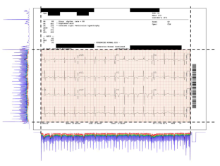
Introduction: T-wave analysis from standard electrocardiogram (ECG) remains one of the most available clinical and research methods for evaluating myocardial repolarization. T-wave morphology was recently evaluated to aid with diagnosis and characterization of diastolic dysfunction. Unfortunately, PDF stored ECG datasets limit additional numerical post-processing of ECG waveforms. In this study, we apply a simple custom process pipeline to extract and re-digitize T-wave signals and subject them to principal component analysis (PCA) to define primary T-wave shape variations.
Methods: We propose simple pre-processing and digitization algorithms programmable as a MATLAB tool using standard thresholding functions without the need for advanced signal analysis. To validate digitized datasets, we compared clinically standard measurements in 20 different ECGs with the original ECG machine interpreted values as a gold standard. Afterwards, we analyzed 212 individual ECGs for T-wave shape analysis using PCA.
Results: The re-digitized signal was shown to preserve the original information as evidenced by excellent agreement between original - machine interpreted and re-digitized clinical variables including heart rate: bias ~ 1 bpm (95% CI: -1.0 to 3.5), QT interval: bias ~ 0.000 ms (95% CI: -0.012 to 0.012), PR interval: bias = -0.015 ms (95% CI: -0.015 to 0.003), and QRS duration: bias = -0.001 ms (95% CI: -0.007 to 0.006). PCA revealed that the first principal component universally modulates the T-wave height or amount of repolarization voltage regardless of the investigated ECG lead. The second and third principal components described variation in the T-wave peak onset and the T-wave peak morphology, respectively.
Conclusion: This study presents a straightforward method for re-digitizing ECGs stored in the PDF format utilized in many academic electronic medical record systems. This process can yield re-digitized lead specific signals which can be retrospectively analyzed using advanced custom post-processing numerical analysis independent of commercially available platforms.
期刊介绍:
Cardiovascular Engineering and Technology is a journal publishing the spectrum of basic to translational research in all aspects of cardiovascular physiology and medical treatment. It is the forum for academic and industrial investigators to disseminate research that utilizes engineering principles and methods to advance fundamental knowledge and technological solutions related to the cardiovascular system. Manuscripts spanning from subcellular to systems level topics are invited, including but not limited to implantable medical devices, hemodynamics and tissue biomechanics, functional imaging, surgical devices, electrophysiology, tissue engineering and regenerative medicine, diagnostic instruments, transport and delivery of biologics, and sensors. In addition to manuscripts describing the original publication of research, manuscripts reviewing developments in these topics or their state-of-art are also invited.

 求助内容:
求助内容: 应助结果提醒方式:


