Kun Zhang, WenBin Ge, ShiTong Luo, Zhi Zhou, YaLi Liu
{"title":"静态磁场促进牙周韧带干细胞的增殖、迁移、分化和AKT激活。","authors":"Kun Zhang, WenBin Ge, ShiTong Luo, Zhi Zhou, YaLi Liu","doi":"10.1159/000524291","DOIUrl":null,"url":null,"abstract":"<p><p>Periodontal ligament stem cells (PDLSCs) possess self-renewal and multilineage differentiation potential and exhibit great potential for the treatment of bone tissue defects caused by inflammation. Previous studies have indicated that static magnetic field (SMF) can enhance the proliferation and differentiation of mesenchymal stem cells (MSCs). SMF has been widely used to repair bone defects and for orthodontic and implantation treatment. In this study, we revealed that a 320 mT SMF upregulates the protein expression levels of cytokines such as MCM7 and PCNA in proliferating PDLSCs. Cell counting kit-8 results revealed that the SMF group had higher optical density values than the control group. The ratio of cells in the S phase to those in the G2/M phase was significantly increased after exposure to a 320 mT SMF. In scratch assays, the SMF-treated PDLSCs exhibited a higher migration rate than the sham-exposed group after 24 h of culture, indicating that the SMF promoted the migratory ability of PDLSCs. The activity level of the early differentiation marker alkaline phosphatase and the late marker matrix mineralization, as well as osteoblast-specific gene and protein expression, were enhanced in PDLSCs exposed to the SMF. Furthermore, AKT signaling pathway was activated by SMF. Our data demonstrated that the potential mechanism of action of SMF may enhance PDLSCs proliferation and osteogenic differentiation by activating the phosphorylated AKT pathway. The elucidation of this molecular mechanism may lead to a better understanding of bone repair responses and aid in improved stem cell-mediated regeneration.</p>","PeriodicalId":9717,"journal":{"name":"Cells Tissues Organs","volume":"212 4","pages":"317-326"},"PeriodicalIF":1.9000,"publicationDate":"2023-01-01","publicationTypes":"Journal Article","fieldsOfStudy":null,"isOpenAccess":false,"openAccessPdf":"https://www.ncbi.nlm.nih.gov/pmc/articles/PMC10534995/pdf/","citationCount":"3","resultStr":"{\"title\":\"Static Magnetic Field Promotes Proliferation, Migration, Differentiation, and AKT Activation of Periodontal Ligament Stem Cells.\",\"authors\":\"Kun Zhang, WenBin Ge, ShiTong Luo, Zhi Zhou, YaLi Liu\",\"doi\":\"10.1159/000524291\",\"DOIUrl\":null,\"url\":null,\"abstract\":\"<p><p>Periodontal ligament stem cells (PDLSCs) possess self-renewal and multilineage differentiation potential and exhibit great potential for the treatment of bone tissue defects caused by inflammation. Previous studies have indicated that static magnetic field (SMF) can enhance the proliferation and differentiation of mesenchymal stem cells (MSCs). SMF has been widely used to repair bone defects and for orthodontic and implantation treatment. In this study, we revealed that a 320 mT SMF upregulates the protein expression levels of cytokines such as MCM7 and PCNA in proliferating PDLSCs. Cell counting kit-8 results revealed that the SMF group had higher optical density values than the control group. The ratio of cells in the S phase to those in the G2/M phase was significantly increased after exposure to a 320 mT SMF. In scratch assays, the SMF-treated PDLSCs exhibited a higher migration rate than the sham-exposed group after 24 h of culture, indicating that the SMF promoted the migratory ability of PDLSCs. The activity level of the early differentiation marker alkaline phosphatase and the late marker matrix mineralization, as well as osteoblast-specific gene and protein expression, were enhanced in PDLSCs exposed to the SMF. Furthermore, AKT signaling pathway was activated by SMF. Our data demonstrated that the potential mechanism of action of SMF may enhance PDLSCs proliferation and osteogenic differentiation by activating the phosphorylated AKT pathway. The elucidation of this molecular mechanism may lead to a better understanding of bone repair responses and aid in improved stem cell-mediated regeneration.</p>\",\"PeriodicalId\":9717,\"journal\":{\"name\":\"Cells Tissues Organs\",\"volume\":\"212 4\",\"pages\":\"317-326\"},\"PeriodicalIF\":1.9000,\"publicationDate\":\"2023-01-01\",\"publicationTypes\":\"Journal Article\",\"fieldsOfStudy\":null,\"isOpenAccess\":false,\"openAccessPdf\":\"https://www.ncbi.nlm.nih.gov/pmc/articles/PMC10534995/pdf/\",\"citationCount\":\"3\",\"resultStr\":null,\"platform\":\"Semanticscholar\",\"paperid\":null,\"PeriodicalName\":\"Cells Tissues Organs\",\"FirstCategoryId\":\"99\",\"ListUrlMain\":\"https://doi.org/10.1159/000524291\",\"RegionNum\":4,\"RegionCategory\":\"生物学\",\"ArticlePicture\":[],\"TitleCN\":null,\"AbstractTextCN\":null,\"PMCID\":null,\"EPubDate\":\"2022/3/28 0:00:00\",\"PubModel\":\"Epub\",\"JCR\":\"Q1\",\"JCRName\":\"ANATOMY & MORPHOLOGY\",\"Score\":null,\"Total\":0}","platform":"Semanticscholar","paperid":null,"PeriodicalName":"Cells Tissues Organs","FirstCategoryId":"99","ListUrlMain":"https://doi.org/10.1159/000524291","RegionNum":4,"RegionCategory":"生物学","ArticlePicture":[],"TitleCN":null,"AbstractTextCN":null,"PMCID":null,"EPubDate":"2022/3/28 0:00:00","PubModel":"Epub","JCR":"Q1","JCRName":"ANATOMY & MORPHOLOGY","Score":null,"Total":0}
Static Magnetic Field Promotes Proliferation, Migration, Differentiation, and AKT Activation of Periodontal Ligament Stem Cells.
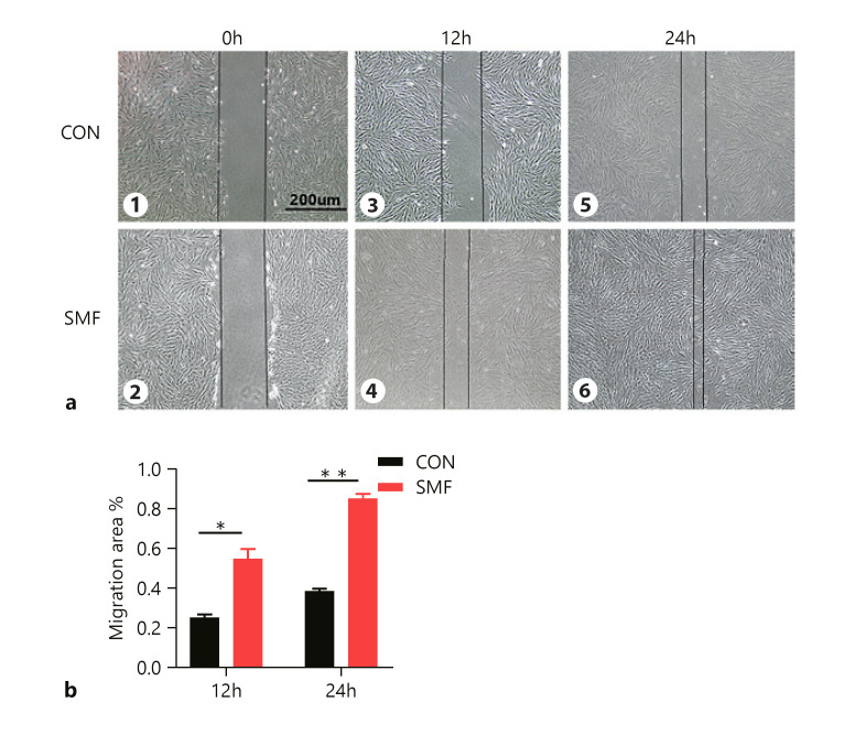
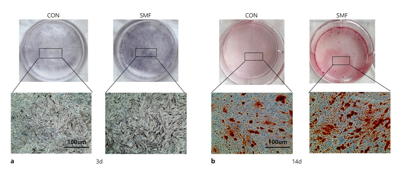
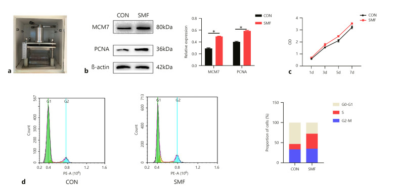
Periodontal ligament stem cells (PDLSCs) possess self-renewal and multilineage differentiation potential and exhibit great potential for the treatment of bone tissue defects caused by inflammation. Previous studies have indicated that static magnetic field (SMF) can enhance the proliferation and differentiation of mesenchymal stem cells (MSCs). SMF has been widely used to repair bone defects and for orthodontic and implantation treatment. In this study, we revealed that a 320 mT SMF upregulates the protein expression levels of cytokines such as MCM7 and PCNA in proliferating PDLSCs. Cell counting kit-8 results revealed that the SMF group had higher optical density values than the control group. The ratio of cells in the S phase to those in the G2/M phase was significantly increased after exposure to a 320 mT SMF. In scratch assays, the SMF-treated PDLSCs exhibited a higher migration rate than the sham-exposed group after 24 h of culture, indicating that the SMF promoted the migratory ability of PDLSCs. The activity level of the early differentiation marker alkaline phosphatase and the late marker matrix mineralization, as well as osteoblast-specific gene and protein expression, were enhanced in PDLSCs exposed to the SMF. Furthermore, AKT signaling pathway was activated by SMF. Our data demonstrated that the potential mechanism of action of SMF may enhance PDLSCs proliferation and osteogenic differentiation by activating the phosphorylated AKT pathway. The elucidation of this molecular mechanism may lead to a better understanding of bone repair responses and aid in improved stem cell-mediated regeneration.
期刊介绍:
''Cells Tissues Organs'' aims at bridging the gap between cell biology and developmental biology and the emerging fields of regenerative medicine (stem cell biology, tissue engineering, artificial organs, in vitro systems and transplantation biology). CTO offers a rapid and fair peer-review and exquisite reproduction quality. Special topic issues, entire issues of the journal devoted to a single research topic within the range of interests of the journal, are published at irregular intervals.
 求助内容:
求助内容: 应助结果提醒方式:


