{"title":"肉芽肿性纵隔炎:肺动脉高压的罕见病因。","authors":"Burcu Öztürk Şahin, Züheyla Galata, Şerife Demir, Nilgün Yılmaz Demirci, Yılmaz Demirci, Gonca Erbaş, İpek Işıl Gönül, Lütfiye Özlem Atay, I Kıvılcım Oğuzülgen","doi":"10.5152/ThoracResPract.2023.22110","DOIUrl":null,"url":null,"abstract":"<p><p>A rare case of a patient with chronic obstructive pulmonary disease who developed secondary anthracofibrosis to biomass exposure, fibrosing mediastinitis due to anthracotic enlarged lymph nodes in the mediastinum, and pulmonary hypertension because of compres- sion of the lymph nodes on the pulmonary arteries is presented. This is a case report of a 71-year-old female patient who has been followed up with chronic obstructive pulmonary disease for 10 years, has no history of smoking, and has been exposed to biomass for many years. The patient, who had been hospitalized in various centers for the last 3 years due to progressive shortness of breath and dry cough, applied to us with dry cough and dyspnea complaints. On echocardiography, systolic pulmonary arterial pressure was found to be 59 mmHg. For the etiology of pulmonary hypertension, dual-energy thoracic computed tomography was performed with the suspicion of chronic thromboembolic pulmonary hypertension. No filling defect compatible with thromboembolism was detected. In right heart catheterization, mean pulmonary artery pressure was 27 mmHg, pulmonary capillary tip pressure was 7 mmHg, and pulmonary vascular resistance was 3.71 woods units. Endobronchial ultrasound was applied to the patient with the preliminary diagnoses of lymphoma, anthracosis, fibrosing mediastinitis, and infection. Widespread anthracosis was observed in all lobes and segments macroscopically. The lymph node in the subcarinal area was interpreted as anthracotic lymph node. Anthracosis is defined as black pigmentation involving the mucosal, and submucosal layers of the tracheobronchial tree and the lung parenchyma. If anthracosis is associated with luminal obliteration and/or mucosal proliferation causing obstruction, it is considered anthracofibrosis. In this case, we saw that secondary anthracofibrosis, fibrosing mediastinitis due to anthracotic enlarged lymph nodes in the mediastinum, and pulmonary hypertension may develop because of compression of the lymph nodes on the pulmonary arteries, and we wanted to draw attention to it was a rare case.</p>","PeriodicalId":75221,"journal":{"name":"Thoracic research and practice","volume":"24 4","pages":"231-234"},"PeriodicalIF":0.0000,"publicationDate":"2023-07-01","publicationTypes":"Journal Article","fieldsOfStudy":null,"isOpenAccess":false,"openAccessPdf":"https://ftp.ncbi.nlm.nih.gov/pub/pmc/oa_pdf/f8/e5/trp-24-4-231.PMC10543070.pdf","citationCount":"0","resultStr":"{\"title\":\"Granulomatous Mediastinitis: A Rare Cause of Pulmonary Hypertension.\",\"authors\":\"Burcu Öztürk Şahin, Züheyla Galata, Şerife Demir, Nilgün Yılmaz Demirci, Yılmaz Demirci, Gonca Erbaş, İpek Işıl Gönül, Lütfiye Özlem Atay, I Kıvılcım Oğuzülgen\",\"doi\":\"10.5152/ThoracResPract.2023.22110\",\"DOIUrl\":null,\"url\":null,\"abstract\":\"<p><p>A rare case of a patient with chronic obstructive pulmonary disease who developed secondary anthracofibrosis to biomass exposure, fibrosing mediastinitis due to anthracotic enlarged lymph nodes in the mediastinum, and pulmonary hypertension because of compres- sion of the lymph nodes on the pulmonary arteries is presented. This is a case report of a 71-year-old female patient who has been followed up with chronic obstructive pulmonary disease for 10 years, has no history of smoking, and has been exposed to biomass for many years. The patient, who had been hospitalized in various centers for the last 3 years due to progressive shortness of breath and dry cough, applied to us with dry cough and dyspnea complaints. On echocardiography, systolic pulmonary arterial pressure was found to be 59 mmHg. For the etiology of pulmonary hypertension, dual-energy thoracic computed tomography was performed with the suspicion of chronic thromboembolic pulmonary hypertension. No filling defect compatible with thromboembolism was detected. In right heart catheterization, mean pulmonary artery pressure was 27 mmHg, pulmonary capillary tip pressure was 7 mmHg, and pulmonary vascular resistance was 3.71 woods units. Endobronchial ultrasound was applied to the patient with the preliminary diagnoses of lymphoma, anthracosis, fibrosing mediastinitis, and infection. Widespread anthracosis was observed in all lobes and segments macroscopically. The lymph node in the subcarinal area was interpreted as anthracotic lymph node. Anthracosis is defined as black pigmentation involving the mucosal, and submucosal layers of the tracheobronchial tree and the lung parenchyma. If anthracosis is associated with luminal obliteration and/or mucosal proliferation causing obstruction, it is considered anthracofibrosis. In this case, we saw that secondary anthracofibrosis, fibrosing mediastinitis due to anthracotic enlarged lymph nodes in the mediastinum, and pulmonary hypertension may develop because of compression of the lymph nodes on the pulmonary arteries, and we wanted to draw attention to it was a rare case.</p>\",\"PeriodicalId\":75221,\"journal\":{\"name\":\"Thoracic research and practice\",\"volume\":\"24 4\",\"pages\":\"231-234\"},\"PeriodicalIF\":0.0000,\"publicationDate\":\"2023-07-01\",\"publicationTypes\":\"Journal Article\",\"fieldsOfStudy\":null,\"isOpenAccess\":false,\"openAccessPdf\":\"https://ftp.ncbi.nlm.nih.gov/pub/pmc/oa_pdf/f8/e5/trp-24-4-231.PMC10543070.pdf\",\"citationCount\":\"0\",\"resultStr\":null,\"platform\":\"Semanticscholar\",\"paperid\":null,\"PeriodicalName\":\"Thoracic research and practice\",\"FirstCategoryId\":\"1085\",\"ListUrlMain\":\"https://doi.org/10.5152/ThoracResPract.2023.22110\",\"RegionNum\":0,\"RegionCategory\":null,\"ArticlePicture\":[],\"TitleCN\":null,\"AbstractTextCN\":null,\"PMCID\":null,\"EPubDate\":\"\",\"PubModel\":\"\",\"JCR\":\"0\",\"JCRName\":\"RESPIRATORY SYSTEM\",\"Score\":null,\"Total\":0}","platform":"Semanticscholar","paperid":null,"PeriodicalName":"Thoracic research and practice","FirstCategoryId":"1085","ListUrlMain":"https://doi.org/10.5152/ThoracResPract.2023.22110","RegionNum":0,"RegionCategory":null,"ArticlePicture":[],"TitleCN":null,"AbstractTextCN":null,"PMCID":null,"EPubDate":"","PubModel":"","JCR":"0","JCRName":"RESPIRATORY SYSTEM","Score":null,"Total":0}
Granulomatous Mediastinitis: A Rare Cause of Pulmonary Hypertension.
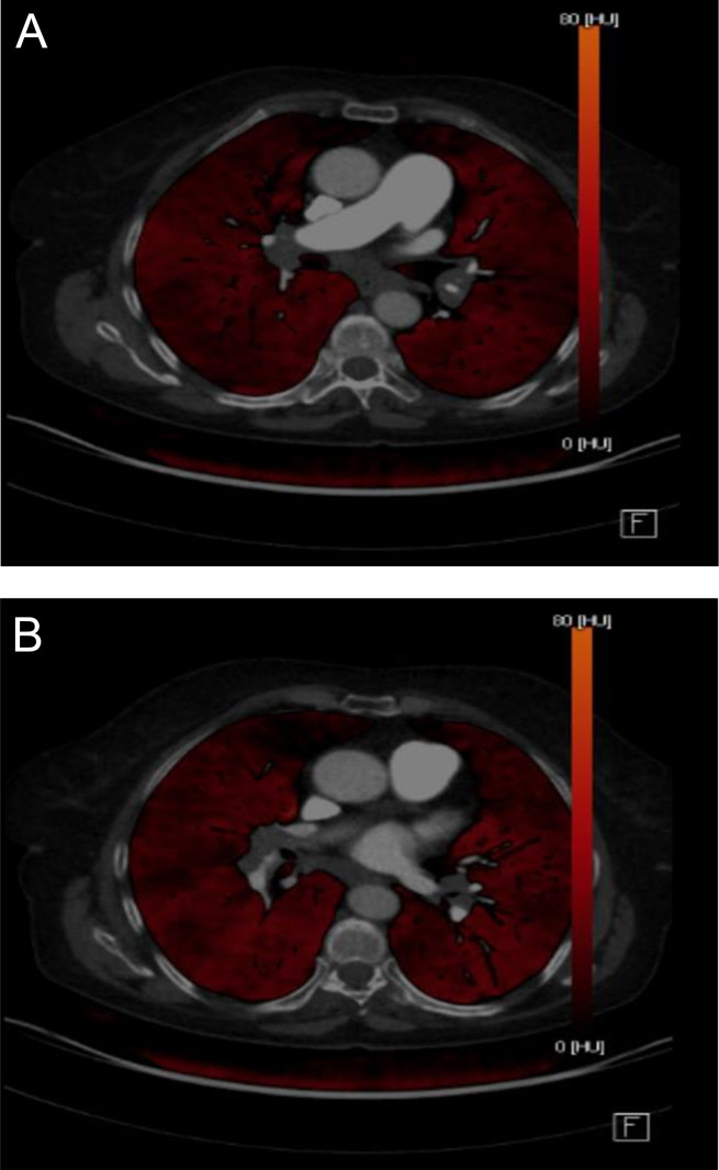
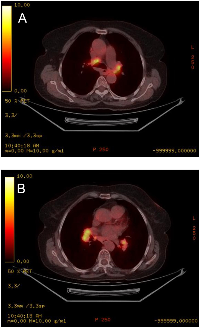
A rare case of a patient with chronic obstructive pulmonary disease who developed secondary anthracofibrosis to biomass exposure, fibrosing mediastinitis due to anthracotic enlarged lymph nodes in the mediastinum, and pulmonary hypertension because of compres- sion of the lymph nodes on the pulmonary arteries is presented. This is a case report of a 71-year-old female patient who has been followed up with chronic obstructive pulmonary disease for 10 years, has no history of smoking, and has been exposed to biomass for many years. The patient, who had been hospitalized in various centers for the last 3 years due to progressive shortness of breath and dry cough, applied to us with dry cough and dyspnea complaints. On echocardiography, systolic pulmonary arterial pressure was found to be 59 mmHg. For the etiology of pulmonary hypertension, dual-energy thoracic computed tomography was performed with the suspicion of chronic thromboembolic pulmonary hypertension. No filling defect compatible with thromboembolism was detected. In right heart catheterization, mean pulmonary artery pressure was 27 mmHg, pulmonary capillary tip pressure was 7 mmHg, and pulmonary vascular resistance was 3.71 woods units. Endobronchial ultrasound was applied to the patient with the preliminary diagnoses of lymphoma, anthracosis, fibrosing mediastinitis, and infection. Widespread anthracosis was observed in all lobes and segments macroscopically. The lymph node in the subcarinal area was interpreted as anthracotic lymph node. Anthracosis is defined as black pigmentation involving the mucosal, and submucosal layers of the tracheobronchial tree and the lung parenchyma. If anthracosis is associated with luminal obliteration and/or mucosal proliferation causing obstruction, it is considered anthracofibrosis. In this case, we saw that secondary anthracofibrosis, fibrosing mediastinitis due to anthracotic enlarged lymph nodes in the mediastinum, and pulmonary hypertension may develop because of compression of the lymph nodes on the pulmonary arteries, and we wanted to draw attention to it was a rare case.


 求助内容:
求助内容: 应助结果提醒方式:


