Antoine Chatrenet, Giorgina Piccoli, Jean Michel Audebrand, Massimo Torreggiani, Julien Barbieux, Charly Vaillant, Baptiste Morel, Sylvain Durand, Bruno Beaune
{"title":"对力量发展率的分析显示,患有慢性肾脏疾病的老年患者具有较高的神经肌肉疲劳能力。","authors":"Antoine Chatrenet, Giorgina Piccoli, Jean Michel Audebrand, Massimo Torreggiani, Julien Barbieux, Charly Vaillant, Baptiste Morel, Sylvain Durand, Bruno Beaune","doi":"10.1002/jcsm.13280","DOIUrl":null,"url":null,"abstract":"<div>\n \n \n <section>\n \n <h3> Background</h3>\n \n <p>Chronic kidney disease (CKD) induces muscle wasting and a reduction in the maximum voluntary force (MVF). Little is known about the neuromuscular fatigability in CKD patients, defined as the reduction of muscle force capacities during exercise. Neuromuscular fatigability is a crucial physical parameter of the daily living. The quantification of explosive force has been shown to be a sensitive means to assess neuromuscular fatigability. Thus, our study used explosive force estimates to assess neuromuscular fatigability in elderly CKD patients.</p>\n </section>\n \n <section>\n \n <h3> Methods</h3>\n \n <p>Inclusion criteria for CKD patients were age ≥ 60 years old and glomerular filtration rate (GFR) < 45 mL/min/1.73 m<sup>2</sup> not on dialysis, and those for controls were GFR > 60 mL/min/1.73 m<sup>2</sup>, age and diabetes matched. The fatigability protocol focused on a handgrip task coupled with surface electromyography (sEMG). Scalars were extracted from the rate of force development (RFD): absolute and normalized time periods (50, 75, 100, 150 and 200 ms, RFD<sub>50</sub>, RFD<sub>75</sub>, RFD<sub>100</sub>, RFD<sub>150</sub> and RFD<sub>200</sub>, respectively), peak RFD (RFD<sub>peak</sub> in absolute; NRFD<sub>peak</sub> normalized), time-to-peak RFD (t-RFD<sub>peak</sub>) and the relative force at RFD<sub>peak</sub> (MVF-RFD<sub>peak</sub>). A statistical parametric mapping approach was performed on the force, impulse and RFD–time curves. The integrated sEMG with time at 0–30, 0–50, 0–100 and 0–200 ms time intervals relative to onset of sEMG activity was extracted and groups were compared separately for each sex.</p>\n </section>\n \n <section>\n \n <h3> Results</h3>\n \n <p>The cohort of 159 individuals had a median age of 69 (9<sub>IQR</sub>) years and body mass index was 27.6 (6.2<sub>IQR</sub>) kg/m<sup>2</sup>. Propensity-score-matched groups balanced CKD patients and controls by gender with 66 males and 34 females. In scalar analysis, CKD patients manifested a higher decrement than controls in the early phase of contraction, regarding the NRFD<sub>peak</sub> (<i>P</i> = 0.009; η<sup>2</sup><sub>p</sub> = 0.034) and RFD<sub>75</sub> and RFD<sub>100</sub> (for both <i>P</i> < 0.001; η<sup>2</sup><sub>p</sub> = 0.068 and 0.064). The one-dimensional analysis confirmed that CKD males manifest higher and delayed neuromuscular fatigability, especially before 100 ms from onset of contraction. sEMG was lower in CKD patients than controls in the 0–100 ms (at rest: <i>P</i> = 0.049, Cohen's <i>d</i> = 0.458) and 0–200 ms (at rest: <i>P</i> = 0.016, Cohen's <i>d</i> = 0.496; during exercise: <i>P</i> = 0.006, Cohen's <i>d</i> = 0.421) time windows. Controls showed greater decrease of sEMG than CKD patients in the 0–30 ms (<i>P</i> = 0.020, Cohen's <i>d</i> = 0.533) and 0–50 ms (<i>P</i> = 0.010, Cohen's <i>d</i> = 0.640) time windows. As opposite to females, males showed almost the same differences between groups.</p>\n </section>\n \n <section>\n \n <h3> Conclusions</h3>\n \n <p>Our study is the first to show that CKD patients have higher fatigability than controls, which may be associated with an impaired motor-unit recruitment, highlighting a neural drive disturbance with CKD. Further studies are needed to confirm these findings.</p>\n </section>\n </div>","PeriodicalId":186,"journal":{"name":"Journal of Cachexia, Sarcopenia and Muscle","volume":"14 5","pages":"2016-2028"},"PeriodicalIF":8.9000,"publicationDate":"2023-07-13","publicationTypes":"Journal Article","fieldsOfStudy":null,"isOpenAccess":false,"openAccessPdf":"https://onlinelibrary.wiley.com/doi/epdf/10.1002/jcsm.13280","citationCount":"1","resultStr":"{\"title\":\"Analysis of the rate of force development reveals high neuromuscular fatigability in elderly patients with chronic kidney disease\",\"authors\":\"Antoine Chatrenet, Giorgina Piccoli, Jean Michel Audebrand, Massimo Torreggiani, Julien Barbieux, Charly Vaillant, Baptiste Morel, Sylvain Durand, Bruno Beaune\",\"doi\":\"10.1002/jcsm.13280\",\"DOIUrl\":null,\"url\":null,\"abstract\":\"<div>\\n \\n \\n <section>\\n \\n <h3> Background</h3>\\n \\n <p>Chronic kidney disease (CKD) induces muscle wasting and a reduction in the maximum voluntary force (MVF). Little is known about the neuromuscular fatigability in CKD patients, defined as the reduction of muscle force capacities during exercise. Neuromuscular fatigability is a crucial physical parameter of the daily living. The quantification of explosive force has been shown to be a sensitive means to assess neuromuscular fatigability. Thus, our study used explosive force estimates to assess neuromuscular fatigability in elderly CKD patients.</p>\\n </section>\\n \\n <section>\\n \\n <h3> Methods</h3>\\n \\n <p>Inclusion criteria for CKD patients were age ≥ 60 years old and glomerular filtration rate (GFR) < 45 mL/min/1.73 m<sup>2</sup> not on dialysis, and those for controls were GFR > 60 mL/min/1.73 m<sup>2</sup>, age and diabetes matched. The fatigability protocol focused on a handgrip task coupled with surface electromyography (sEMG). Scalars were extracted from the rate of force development (RFD): absolute and normalized time periods (50, 75, 100, 150 and 200 ms, RFD<sub>50</sub>, RFD<sub>75</sub>, RFD<sub>100</sub>, RFD<sub>150</sub> and RFD<sub>200</sub>, respectively), peak RFD (RFD<sub>peak</sub> in absolute; NRFD<sub>peak</sub> normalized), time-to-peak RFD (t-RFD<sub>peak</sub>) and the relative force at RFD<sub>peak</sub> (MVF-RFD<sub>peak</sub>). A statistical parametric mapping approach was performed on the force, impulse and RFD–time curves. The integrated sEMG with time at 0–30, 0–50, 0–100 and 0–200 ms time intervals relative to onset of sEMG activity was extracted and groups were compared separately for each sex.</p>\\n </section>\\n \\n <section>\\n \\n <h3> Results</h3>\\n \\n <p>The cohort of 159 individuals had a median age of 69 (9<sub>IQR</sub>) years and body mass index was 27.6 (6.2<sub>IQR</sub>) kg/m<sup>2</sup>. Propensity-score-matched groups balanced CKD patients and controls by gender with 66 males and 34 females. In scalar analysis, CKD patients manifested a higher decrement than controls in the early phase of contraction, regarding the NRFD<sub>peak</sub> (<i>P</i> = 0.009; η<sup>2</sup><sub>p</sub> = 0.034) and RFD<sub>75</sub> and RFD<sub>100</sub> (for both <i>P</i> < 0.001; η<sup>2</sup><sub>p</sub> = 0.068 and 0.064). The one-dimensional analysis confirmed that CKD males manifest higher and delayed neuromuscular fatigability, especially before 100 ms from onset of contraction. sEMG was lower in CKD patients than controls in the 0–100 ms (at rest: <i>P</i> = 0.049, Cohen's <i>d</i> = 0.458) and 0–200 ms (at rest: <i>P</i> = 0.016, Cohen's <i>d</i> = 0.496; during exercise: <i>P</i> = 0.006, Cohen's <i>d</i> = 0.421) time windows. Controls showed greater decrease of sEMG than CKD patients in the 0–30 ms (<i>P</i> = 0.020, Cohen's <i>d</i> = 0.533) and 0–50 ms (<i>P</i> = 0.010, Cohen's <i>d</i> = 0.640) time windows. As opposite to females, males showed almost the same differences between groups.</p>\\n </section>\\n \\n <section>\\n \\n <h3> Conclusions</h3>\\n \\n <p>Our study is the first to show that CKD patients have higher fatigability than controls, which may be associated with an impaired motor-unit recruitment, highlighting a neural drive disturbance with CKD. Further studies are needed to confirm these findings.</p>\\n </section>\\n </div>\",\"PeriodicalId\":186,\"journal\":{\"name\":\"Journal of Cachexia, Sarcopenia and Muscle\",\"volume\":\"14 5\",\"pages\":\"2016-2028\"},\"PeriodicalIF\":8.9000,\"publicationDate\":\"2023-07-13\",\"publicationTypes\":\"Journal Article\",\"fieldsOfStudy\":null,\"isOpenAccess\":false,\"openAccessPdf\":\"https://onlinelibrary.wiley.com/doi/epdf/10.1002/jcsm.13280\",\"citationCount\":\"1\",\"resultStr\":null,\"platform\":\"Semanticscholar\",\"paperid\":null,\"PeriodicalName\":\"Journal of Cachexia, Sarcopenia and Muscle\",\"FirstCategoryId\":\"3\",\"ListUrlMain\":\"https://onlinelibrary.wiley.com/doi/10.1002/jcsm.13280\",\"RegionNum\":1,\"RegionCategory\":\"医学\",\"ArticlePicture\":[],\"TitleCN\":null,\"AbstractTextCN\":null,\"PMCID\":null,\"EPubDate\":\"\",\"PubModel\":\"\",\"JCR\":\"\",\"JCRName\":\"\",\"Score\":null,\"Total\":0}","platform":"Semanticscholar","paperid":null,"PeriodicalName":"Journal of Cachexia, Sarcopenia and Muscle","FirstCategoryId":"3","ListUrlMain":"https://onlinelibrary.wiley.com/doi/10.1002/jcsm.13280","RegionNum":1,"RegionCategory":"医学","ArticlePicture":[],"TitleCN":null,"AbstractTextCN":null,"PMCID":null,"EPubDate":"","PubModel":"","JCR":"","JCRName":"","Score":null,"Total":0}
Analysis of the rate of force development reveals high neuromuscular fatigability in elderly patients with chronic kidney disease
Background
Chronic kidney disease (CKD) induces muscle wasting and a reduction in the maximum voluntary force (MVF). Little is known about the neuromuscular fatigability in CKD patients, defined as the reduction of muscle force capacities during exercise. Neuromuscular fatigability is a crucial physical parameter of the daily living. The quantification of explosive force has been shown to be a sensitive means to assess neuromuscular fatigability. Thus, our study used explosive force estimates to assess neuromuscular fatigability in elderly CKD patients.
Methods
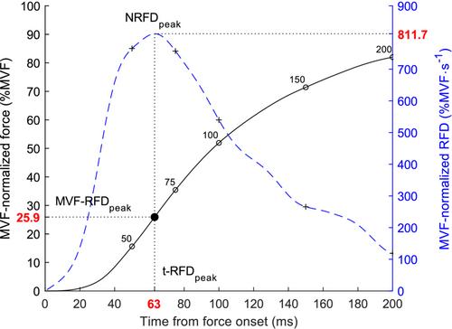
Inclusion criteria for CKD patients were age ≥ 60 years old and glomerular filtration rate (GFR) < 45 mL/min/1.73 m2 not on dialysis, and those for controls were GFR > 60 mL/min/1.73 m2, age and diabetes matched. The fatigability protocol focused on a handgrip task coupled with surface electromyography (sEMG). Scalars were extracted from the rate of force development (RFD): absolute and normalized time periods (50, 75, 100, 150 and 200 ms, RFD50, RFD75, RFD100, RFD150 and RFD200, respectively), peak RFD (RFDpeak in absolute; NRFDpeak normalized), time-to-peak RFD (t-RFDpeak) and the relative force at RFDpeak (MVF-RFDpeak). A statistical parametric mapping approach was performed on the force, impulse and RFD–time curves. The integrated sEMG with time at 0–30, 0–50, 0–100 and 0–200 ms time intervals relative to onset of sEMG activity was extracted and groups were compared separately for each sex.
Results
The cohort of 159 individuals had a median age of 69 (9IQR) years and body mass index was 27.6 (6.2IQR) kg/m2. Propensity-score-matched groups balanced CKD patients and controls by gender with 66 males and 34 females. In scalar analysis, CKD patients manifested a higher decrement than controls in the early phase of contraction, regarding the NRFDpeak (P = 0.009; η2p = 0.034) and RFD75 and RFD100 (for both P < 0.001; η2p = 0.068 and 0.064). The one-dimensional analysis confirmed that CKD males manifest higher and delayed neuromuscular fatigability, especially before 100 ms from onset of contraction. sEMG was lower in CKD patients than controls in the 0–100 ms (at rest: P = 0.049, Cohen's d = 0.458) and 0–200 ms (at rest: P = 0.016, Cohen's d = 0.496; during exercise: P = 0.006, Cohen's d = 0.421) time windows. Controls showed greater decrease of sEMG than CKD patients in the 0–30 ms (P = 0.020, Cohen's d = 0.533) and 0–50 ms (P = 0.010, Cohen's d = 0.640) time windows. As opposite to females, males showed almost the same differences between groups.
Conclusions
Our study is the first to show that CKD patients have higher fatigability than controls, which may be associated with an impaired motor-unit recruitment, highlighting a neural drive disturbance with CKD. Further studies are needed to confirm these findings.
期刊介绍:
The Journal of Cachexia, Sarcopenia, and Muscle is a prestigious, peer-reviewed international publication committed to disseminating research and clinical insights pertaining to cachexia, sarcopenia, body composition, and the physiological and pathophysiological alterations occurring throughout the lifespan and in various illnesses across the spectrum of life sciences. This journal serves as a valuable resource for physicians, biochemists, biologists, dieticians, pharmacologists, and students alike.
 求助内容:
求助内容: 应助结果提醒方式:


