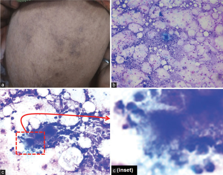
{"title":"乳房肿块:“让我加入你的差异”。","authors":"Manjit Kaur, Neelam Chaudhary, Gargi Kapatia","doi":"10.25259/Cytojournal_12_2022","DOIUrl":null,"url":null,"abstract":"1. What is the diagnosis based on cytomorphology? a. Actinomyces b. Tuberculosis c. Non-Hodgkin’s lymphoma d. Ductal carcinoma. Figure 1: a. Left breast with erythema. (b,c, and c (inset). FNA smear showing inflammatory cells comprising of neutrophils, foamy macrophages, and degenerated cells along with cotton ball like colony in the center with radiating filaments [b, c, and c(inset): MGG; b, X10; c, X40; c(inset) Zoomed]. b","PeriodicalId":49082,"journal":{"name":"Cytojournal","volume":null,"pages":null},"PeriodicalIF":2.5000,"publicationDate":"2023-01-01","publicationTypes":"Journal Article","fieldsOfStudy":null,"isOpenAccess":false,"openAccessPdf":"https://www.ncbi.nlm.nih.gov/pmc/articles/PMC10316205/pdf/","citationCount":"0","resultStr":"{\"title\":\"Breast lump: \\\"Keep me in your differentials\\\".\",\"authors\":\"Manjit Kaur, Neelam Chaudhary, Gargi Kapatia\",\"doi\":\"10.25259/Cytojournal_12_2022\",\"DOIUrl\":null,\"url\":null,\"abstract\":\"1. What is the diagnosis based on cytomorphology? a. Actinomyces b. Tuberculosis c. Non-Hodgkin’s lymphoma d. Ductal carcinoma. Figure 1: a. Left breast with erythema. (b,c, and c (inset). FNA smear showing inflammatory cells comprising of neutrophils, foamy macrophages, and degenerated cells along with cotton ball like colony in the center with radiating filaments [b, c, and c(inset): MGG; b, X10; c, X40; c(inset) Zoomed]. b\",\"PeriodicalId\":49082,\"journal\":{\"name\":\"Cytojournal\",\"volume\":null,\"pages\":null},\"PeriodicalIF\":2.5000,\"publicationDate\":\"2023-01-01\",\"publicationTypes\":\"Journal Article\",\"fieldsOfStudy\":null,\"isOpenAccess\":false,\"openAccessPdf\":\"https://www.ncbi.nlm.nih.gov/pmc/articles/PMC10316205/pdf/\",\"citationCount\":\"0\",\"resultStr\":null,\"platform\":\"Semanticscholar\",\"paperid\":null,\"PeriodicalName\":\"Cytojournal\",\"FirstCategoryId\":\"3\",\"ListUrlMain\":\"https://doi.org/10.25259/Cytojournal_12_2022\",\"RegionNum\":4,\"RegionCategory\":\"医学\",\"ArticlePicture\":[],\"TitleCN\":null,\"AbstractTextCN\":null,\"PMCID\":null,\"EPubDate\":\"\",\"PubModel\":\"\",\"JCR\":\"Q2\",\"JCRName\":\"PATHOLOGY\",\"Score\":null,\"Total\":0}","platform":"Semanticscholar","paperid":null,"PeriodicalName":"Cytojournal","FirstCategoryId":"3","ListUrlMain":"https://doi.org/10.25259/Cytojournal_12_2022","RegionNum":4,"RegionCategory":"医学","ArticlePicture":[],"TitleCN":null,"AbstractTextCN":null,"PMCID":null,"EPubDate":"","PubModel":"","JCR":"Q2","JCRName":"PATHOLOGY","Score":null,"Total":0}
1. What is the diagnosis based on cytomorphology? a. Actinomyces b. Tuberculosis c. Non-Hodgkin’s lymphoma d. Ductal carcinoma. Figure 1: a. Left breast with erythema. (b,c, and c (inset). FNA smear showing inflammatory cells comprising of neutrophils, foamy macrophages, and degenerated cells along with cotton ball like colony in the center with radiating filaments [b, c, and c(inset): MGG; b, X10; c, X40; c(inset) Zoomed]. b
期刊介绍:
The CytoJournal is an open-access peer-reviewed journal committed to publishing high-quality articles in the field of Diagnostic Cytopathology including Molecular aspects. The journal is owned by the Cytopathology Foundation and published by the Scientific Scholar.