{"title":"两种血小板浓缩物(a-PRF和L-PRF)对成骨前MG-63细胞活性影响的体外比较研究","authors":"Azadeh Esmaeilnejad, Mohammadreza Talebi Ardakani, Mahdi Shokri, PhD Nima Hosseini Khou, Mobina Kamani","doi":"10.30476/dentjods.2022.93305.1709","DOIUrl":null,"url":null,"abstract":"<p><strong>Statement of the problem: </strong>Currently, the reconstruction of bone defects with new platelet concentrates is considered a significant challenge in periodontics.</p><p><strong>Purpose: </strong>This study aimed to evaluate advanced- platelet rich fibrin (A-PRF) and leukocyte- and platelet rich fibrin's (L-PRF) effects on the proliferation and differentiation of MG-63 cells.</p><p><strong>Materials and method: </strong>In this <i>in vitro</i> study, blood samples of five healthy non-smoking volunteers were collected and immediately centrifuged according to the two protocols of Choukroun and Ghanaati, without adding any anticoagulants, to prepare L-PRF and A-PRF. After freezing the clots for one hour, they were crushed and centrifuged once more. After culturing MG-63 cells, the effects of 20%, 10%, 1%, and 0.5% concentrations of A-PRF and L-PRF extracts on cell proliferation and mineralization were evaluated by methyl thiazolyl tetrazolium (MTT) assay and Alizarin Red staining, respectively.</p><p><strong>Results: </strong>Generally, survival and proliferation in the L-PRF group at both time intervals were higher than the A-PRF group and increased with increasing the extract concentration. However, in the A-PRF group, there were no significant differences between the different concentrations, and only the number of cells increased over time. After three days, in the study on mineralization, nodule formation was observed only in the positive control group (osteogenic). In seven days, mineralized nodules were formed in all groups with different concentrations of A-PRF, but not in any of the L-PRF groups.</p><p><strong>Conclusion: </strong>According to the results, L-PRF increased proliferation, and A-PRF exerted a positive effect on the differentiation of MG-63 cells.</p>","PeriodicalId":73702,"journal":{"name":"Journal of dentistry (Shiraz, Iran)","volume":"24 2","pages":"235-244"},"PeriodicalIF":0.0000,"publicationDate":"2023-06-01","publicationTypes":"Journal Article","fieldsOfStudy":null,"isOpenAccess":false,"openAccessPdf":"https://www.ncbi.nlm.nih.gov/pmc/articles/PMC10300147/pdf/","citationCount":"3","resultStr":"{\"title\":\"Comparative Evaluation of the Effect of Two Platelet Concentrates (a-PRF and L-PRF) on the Cellular Activity of Pre-osteoblastic MG-63 Cell Line: An in vitro Study.\",\"authors\":\"Azadeh Esmaeilnejad, Mohammadreza Talebi Ardakani, Mahdi Shokri, PhD Nima Hosseini Khou, Mobina Kamani\",\"doi\":\"10.30476/dentjods.2022.93305.1709\",\"DOIUrl\":null,\"url\":null,\"abstract\":\"<p><strong>Statement of the problem: </strong>Currently, the reconstruction of bone defects with new platelet concentrates is considered a significant challenge in periodontics.</p><p><strong>Purpose: </strong>This study aimed to evaluate advanced- platelet rich fibrin (A-PRF) and leukocyte- and platelet rich fibrin's (L-PRF) effects on the proliferation and differentiation of MG-63 cells.</p><p><strong>Materials and method: </strong>In this <i>in vitro</i> study, blood samples of five healthy non-smoking volunteers were collected and immediately centrifuged according to the two protocols of Choukroun and Ghanaati, without adding any anticoagulants, to prepare L-PRF and A-PRF. After freezing the clots for one hour, they were crushed and centrifuged once more. After culturing MG-63 cells, the effects of 20%, 10%, 1%, and 0.5% concentrations of A-PRF and L-PRF extracts on cell proliferation and mineralization were evaluated by methyl thiazolyl tetrazolium (MTT) assay and Alizarin Red staining, respectively.</p><p><strong>Results: </strong>Generally, survival and proliferation in the L-PRF group at both time intervals were higher than the A-PRF group and increased with increasing the extract concentration. However, in the A-PRF group, there were no significant differences between the different concentrations, and only the number of cells increased over time. After three days, in the study on mineralization, nodule formation was observed only in the positive control group (osteogenic). In seven days, mineralized nodules were formed in all groups with different concentrations of A-PRF, but not in any of the L-PRF groups.</p><p><strong>Conclusion: </strong>According to the results, L-PRF increased proliferation, and A-PRF exerted a positive effect on the differentiation of MG-63 cells.</p>\",\"PeriodicalId\":73702,\"journal\":{\"name\":\"Journal of dentistry (Shiraz, Iran)\",\"volume\":\"24 2\",\"pages\":\"235-244\"},\"PeriodicalIF\":0.0000,\"publicationDate\":\"2023-06-01\",\"publicationTypes\":\"Journal Article\",\"fieldsOfStudy\":null,\"isOpenAccess\":false,\"openAccessPdf\":\"https://www.ncbi.nlm.nih.gov/pmc/articles/PMC10300147/pdf/\",\"citationCount\":\"3\",\"resultStr\":null,\"platform\":\"Semanticscholar\",\"paperid\":null,\"PeriodicalName\":\"Journal of dentistry (Shiraz, Iran)\",\"FirstCategoryId\":\"1085\",\"ListUrlMain\":\"https://doi.org/10.30476/dentjods.2022.93305.1709\",\"RegionNum\":0,\"RegionCategory\":null,\"ArticlePicture\":[],\"TitleCN\":null,\"AbstractTextCN\":null,\"PMCID\":null,\"EPubDate\":\"\",\"PubModel\":\"\",\"JCR\":\"\",\"JCRName\":\"\",\"Score\":null,\"Total\":0}","platform":"Semanticscholar","paperid":null,"PeriodicalName":"Journal of dentistry (Shiraz, Iran)","FirstCategoryId":"1085","ListUrlMain":"https://doi.org/10.30476/dentjods.2022.93305.1709","RegionNum":0,"RegionCategory":null,"ArticlePicture":[],"TitleCN":null,"AbstractTextCN":null,"PMCID":null,"EPubDate":"","PubModel":"","JCR":"","JCRName":"","Score":null,"Total":0}
Comparative Evaluation of the Effect of Two Platelet Concentrates (a-PRF and L-PRF) on the Cellular Activity of Pre-osteoblastic MG-63 Cell Line: An in vitro Study.
Statement of the problem: Currently, the reconstruction of bone defects with new platelet concentrates is considered a significant challenge in periodontics.
Purpose: This study aimed to evaluate advanced- platelet rich fibrin (A-PRF) and leukocyte- and platelet rich fibrin's (L-PRF) effects on the proliferation and differentiation of MG-63 cells.
Materials and method: In this in vitro study, blood samples of five healthy non-smoking volunteers were collected and immediately centrifuged according to the two protocols of Choukroun and Ghanaati, without adding any anticoagulants, to prepare L-PRF and A-PRF. After freezing the clots for one hour, they were crushed and centrifuged once more. After culturing MG-63 cells, the effects of 20%, 10%, 1%, and 0.5% concentrations of A-PRF and L-PRF extracts on cell proliferation and mineralization were evaluated by methyl thiazolyl tetrazolium (MTT) assay and Alizarin Red staining, respectively.
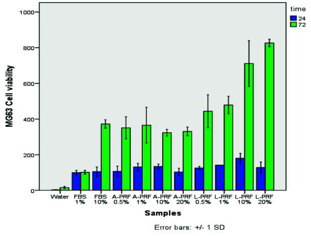
Results: Generally, survival and proliferation in the L-PRF group at both time intervals were higher than the A-PRF group and increased with increasing the extract concentration. However, in the A-PRF group, there were no significant differences between the different concentrations, and only the number of cells increased over time. After three days, in the study on mineralization, nodule formation was observed only in the positive control group (osteogenic). In seven days, mineralized nodules were formed in all groups with different concentrations of A-PRF, but not in any of the L-PRF groups.
Conclusion: According to the results, L-PRF increased proliferation, and A-PRF exerted a positive effect on the differentiation of MG-63 cells.

 求助内容:
求助内容: 应助结果提醒方式:


