Shun-Ichi Kawarai, Shintaro Katahira, Midori Miyatake, Kota Itagaki, Noriko Tsuruoka, Yoichi Haga, Yoshikatsu Saiki
{"title":"开发用于实时监测食管粘膜血流的改良型激光多普勒血流测量仪:通过动物模型进行临床前评估。","authors":"Shun-Ichi Kawarai, Shintaro Katahira, Midori Miyatake, Kota Itagaki, Noriko Tsuruoka, Yoichi Haga, Yoshikatsu Saiki","doi":"10.1007/s10047-023-01408-w","DOIUrl":null,"url":null,"abstract":"<p><p>This study aimed to modify a laser Doppler flowmeter designed and assembled at our institute. After measuring sensitivity evaluation in ex vivo experiments, we confirmed the efficacy of this new device for monitoring real-time esophageal mucosal blood flow changes after thoracic stent graft implantation by simulating various clinical situations in an animal model. Thoracic stent graft implantation was performed in a swine model (n = 8). Esophageal mucosal blood flow decreased significantly from baseline (34.1 ± 18.8 ml/min/100 g vs. 16.7 ± 6.6 ml/min/100 g, P < 0.05) in the lower esophagus (Th6-Th8) where the stent graft covered the aorta. In the hemorrhagic shock model (shock index ≥ 1.0), esophageal mucosal blood flow showed a remarkable change from baseline in the upper esophagus (Th1-Th3), where the stent graft did not cover the aorta (20.8 ± 9.8 ml/min/100 g vs. 12.9 ± 8.6 ml/min/100 g, P < 0.01); however, it returned to the baseline value within a 30-min period. Mucosal blood flow remained stable in the esophagus, where the stent graft did not cover the aorta. After elevating the mean blood pressure to > 70 mmHg with continuous intravenous noradrenaline infusion, esophageal mucosal blood flow increased significantly in both regions; however, the reaction was different between the two regions. Our newly developed laser Doppler flowmeter could measure real-time esophageal mucosal blood flow changes in various clinical situations during thoracic stent graft implantation in a swine model. Hence, this device can be applied in many medical fields by downsizing it.</p>","PeriodicalId":15177,"journal":{"name":"Journal of Artificial Organs","volume":" ","pages":"284-292"},"PeriodicalIF":1.3000,"publicationDate":"2024-09-01","publicationTypes":"Journal Article","fieldsOfStudy":null,"isOpenAccess":false,"openAccessPdf":"","citationCount":"0","resultStr":"{\"title\":\"Development of modified laser Doppler flowmetry device for real-time monitoring of esophageal mucosal blood flow: a preclinical assessment with an animal model.\",\"authors\":\"Shun-Ichi Kawarai, Shintaro Katahira, Midori Miyatake, Kota Itagaki, Noriko Tsuruoka, Yoichi Haga, Yoshikatsu Saiki\",\"doi\":\"10.1007/s10047-023-01408-w\",\"DOIUrl\":null,\"url\":null,\"abstract\":\"<p><p>This study aimed to modify a laser Doppler flowmeter designed and assembled at our institute. After measuring sensitivity evaluation in ex vivo experiments, we confirmed the efficacy of this new device for monitoring real-time esophageal mucosal blood flow changes after thoracic stent graft implantation by simulating various clinical situations in an animal model. Thoracic stent graft implantation was performed in a swine model (n = 8). Esophageal mucosal blood flow decreased significantly from baseline (34.1 ± 18.8 ml/min/100 g vs. 16.7 ± 6.6 ml/min/100 g, P < 0.05) in the lower esophagus (Th6-Th8) where the stent graft covered the aorta. In the hemorrhagic shock model (shock index ≥ 1.0), esophageal mucosal blood flow showed a remarkable change from baseline in the upper esophagus (Th1-Th3), where the stent graft did not cover the aorta (20.8 ± 9.8 ml/min/100 g vs. 12.9 ± 8.6 ml/min/100 g, P < 0.01); however, it returned to the baseline value within a 30-min period. Mucosal blood flow remained stable in the esophagus, where the stent graft did not cover the aorta. After elevating the mean blood pressure to > 70 mmHg with continuous intravenous noradrenaline infusion, esophageal mucosal blood flow increased significantly in both regions; however, the reaction was different between the two regions. Our newly developed laser Doppler flowmeter could measure real-time esophageal mucosal blood flow changes in various clinical situations during thoracic stent graft implantation in a swine model. Hence, this device can be applied in many medical fields by downsizing it.</p>\",\"PeriodicalId\":15177,\"journal\":{\"name\":\"Journal of Artificial Organs\",\"volume\":\" \",\"pages\":\"284-292\"},\"PeriodicalIF\":1.3000,\"publicationDate\":\"2024-09-01\",\"publicationTypes\":\"Journal Article\",\"fieldsOfStudy\":null,\"isOpenAccess\":false,\"openAccessPdf\":\"\",\"citationCount\":\"0\",\"resultStr\":null,\"platform\":\"Semanticscholar\",\"paperid\":null,\"PeriodicalName\":\"Journal of Artificial Organs\",\"FirstCategoryId\":\"5\",\"ListUrlMain\":\"https://doi.org/10.1007/s10047-023-01408-w\",\"RegionNum\":4,\"RegionCategory\":\"医学\",\"ArticlePicture\":[],\"TitleCN\":null,\"AbstractTextCN\":null,\"PMCID\":null,\"EPubDate\":\"2023/7/7 0:00:00\",\"PubModel\":\"Epub\",\"JCR\":\"Q4\",\"JCRName\":\"ENGINEERING, BIOMEDICAL\",\"Score\":null,\"Total\":0}","platform":"Semanticscholar","paperid":null,"PeriodicalName":"Journal of Artificial Organs","FirstCategoryId":"5","ListUrlMain":"https://doi.org/10.1007/s10047-023-01408-w","RegionNum":4,"RegionCategory":"医学","ArticlePicture":[],"TitleCN":null,"AbstractTextCN":null,"PMCID":null,"EPubDate":"2023/7/7 0:00:00","PubModel":"Epub","JCR":"Q4","JCRName":"ENGINEERING, BIOMEDICAL","Score":null,"Total":0}
引用次数: 0
摘要
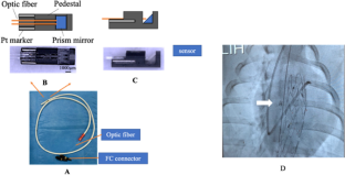
本研究旨在改进本研究所设计和组装的激光多普勒血流计。在体内外实验中进行灵敏度评估后,我们通过在动物模型中模拟各种临床情况,证实了这种新设备在胸腔支架移植物植入后实时监测食管粘膜血流变化的有效性。在猪模型(n = 8)中进行了胸腔支架移植物植入术。食管粘膜血流较基线明显下降(34.1 ± 18.8 ml/min/100 g vs. 16.7 ± 6.6 ml/min/100 g,P 70 mmHg),持续静脉输注去甲肾上腺素后,两个区域的食管粘膜血流均明显增加,但两个区域的反应不同。我们新开发的激光多普勒血流测量仪可在猪模型中实时测量胸腔支架移植过程中各种临床情况下食管粘膜血流的变化。因此,该装置通过小型化可应用于许多医疗领域。
Development of modified laser Doppler flowmetry device for real-time monitoring of esophageal mucosal blood flow: a preclinical assessment with an animal model.
This study aimed to modify a laser Doppler flowmeter designed and assembled at our institute. After measuring sensitivity evaluation in ex vivo experiments, we confirmed the efficacy of this new device for monitoring real-time esophageal mucosal blood flow changes after thoracic stent graft implantation by simulating various clinical situations in an animal model. Thoracic stent graft implantation was performed in a swine model (n = 8). Esophageal mucosal blood flow decreased significantly from baseline (34.1 ± 18.8 ml/min/100 g vs. 16.7 ± 6.6 ml/min/100 g, P < 0.05) in the lower esophagus (Th6-Th8) where the stent graft covered the aorta. In the hemorrhagic shock model (shock index ≥ 1.0), esophageal mucosal blood flow showed a remarkable change from baseline in the upper esophagus (Th1-Th3), where the stent graft did not cover the aorta (20.8 ± 9.8 ml/min/100 g vs. 12.9 ± 8.6 ml/min/100 g, P < 0.01); however, it returned to the baseline value within a 30-min period. Mucosal blood flow remained stable in the esophagus, where the stent graft did not cover the aorta. After elevating the mean blood pressure to > 70 mmHg with continuous intravenous noradrenaline infusion, esophageal mucosal blood flow increased significantly in both regions; however, the reaction was different between the two regions. Our newly developed laser Doppler flowmeter could measure real-time esophageal mucosal blood flow changes in various clinical situations during thoracic stent graft implantation in a swine model. Hence, this device can be applied in many medical fields by downsizing it.
期刊介绍:
The aim of the Journal of Artificial Organs is to introduce to colleagues worldwide a broad spectrum of important new achievements in the field of artificial organs, ranging from fundamental research to clinical applications. The scope of the Journal of Artificial Organs encompasses but is not restricted to blood purification, cardiovascular intervention, biomaterials, and artificial metabolic organs. Additionally, the journal will cover technical and industrial innovations. Membership in the Japanese Society for Artificial Organs is not a prerequisite for submission.

 求助内容:
求助内容: 应助结果提醒方式:


