Dongseob Lee, Jungwon Lee, Ki-Tae Koo, Yang-Jo Seol, Yong-Moo Lee
{"title":"多脱氧核糖核酸对侧窗窦底抬高同时植入早期骨形成的影响。","authors":"Dongseob Lee, Jungwon Lee, Ki-Tae Koo, Yang-Jo Seol, Yong-Moo Lee","doi":"10.5051/jpis.2202760138","DOIUrl":null,"url":null,"abstract":"<p><strong>Purpose: </strong>The aim of this study was to evaluate the impact of polydeoxyribonucleotide (PDRN) on histologic outcomes when implant placement and lateral sinus floor elevation are performed simultaneously.</p><p><strong>Methods: </strong>Three bimaxillary premolars (P2, P3, and P4) were extracted from 4 beagle dogs 2 months before lateral sinus floor elevation. After lateral elevation of the sinus membrane, each sinus was allocated to either the test or control group. Sinuses underwent either 1) collagenated synthetic bone graft with PDRN following lateral sinus floor elevation (test group) or 2) collagenated synthetic bone graft without PDRN after lateral sinus floor elevation (control group). Eight weeks after the surgical procedure, all animals were euthanised for a histologic and histomorphometric assessment. Augmented height (AH), protruding height (PH), and bone-to-implant contact in pristine (BIC<sub>p</sub>) and augmented (BIC<sub>a</sub>) bone were measured. The composition of the augmented area, which was divided into 3 areas of interest located in coronal, middle and apical areas (AOI_C, AOI_M, and AOI_A), was calculated with 3 parameters: the area percentage of new bone (pNB), residual bone graft particle (pRBP), and fibrovascular connective tissue (pFVT).</p><p><strong>Results: </strong>AH, PH, BIC<sub>p</sub>, BIC<sub>a</sub> total, BIC<sub>a</sub> coronal, and BIC<sub>a</sub> middle values were not significantly different between sinuses in the control and test groups (all <i>P</i>>0.05). The BIC<sub>a</sub> apical of sinuses in the test group (76.7%±9.3%) showed statistically higher values than those of sinuses in the control group (55.6%±22.1%) (<i>P</i>=0.038). pNB, pRBP, and pFVT showed statistically significant differences between the 2 groups in AOI_A (<i>P</i>=0.038, <i>P=</i>0.028, and <i>P=</i>0.007, respectively). pNB, pRBP, and pFVT in AOI_C and AOI_M were not significantly different between samples in the control and test groups (all <i>P</i>>0.05).</p><p><strong>Conclusions: </strong>The histologic findings revealed that lateral sinus floor elevation with PDRN might improve early new bone formation and enable higher bone-to-implant contact.</p>","PeriodicalId":48795,"journal":{"name":"Journal of Periodontal and Implant Science","volume":"53 2","pages":"157-169"},"PeriodicalIF":2.2000,"publicationDate":"2023-04-01","publicationTypes":"Journal Article","fieldsOfStudy":null,"isOpenAccess":false,"openAccessPdf":"https://ftp.ncbi.nlm.nih.gov/pub/pmc/oa_pdf/0d/60/jpis-53-157.PMC10133820.pdf","citationCount":"4","resultStr":"{\"title\":\"The impact of polydeoxyribonucleotide on early bone formation in lateral-window sinus floor elevation with simultaneous implant placement.\",\"authors\":\"Dongseob Lee, Jungwon Lee, Ki-Tae Koo, Yang-Jo Seol, Yong-Moo Lee\",\"doi\":\"10.5051/jpis.2202760138\",\"DOIUrl\":null,\"url\":null,\"abstract\":\"<p><strong>Purpose: </strong>The aim of this study was to evaluate the impact of polydeoxyribonucleotide (PDRN) on histologic outcomes when implant placement and lateral sinus floor elevation are performed simultaneously.</p><p><strong>Methods: </strong>Three bimaxillary premolars (P2, P3, and P4) were extracted from 4 beagle dogs 2 months before lateral sinus floor elevation. After lateral elevation of the sinus membrane, each sinus was allocated to either the test or control group. Sinuses underwent either 1) collagenated synthetic bone graft with PDRN following lateral sinus floor elevation (test group) or 2) collagenated synthetic bone graft without PDRN after lateral sinus floor elevation (control group). Eight weeks after the surgical procedure, all animals were euthanised for a histologic and histomorphometric assessment. Augmented height (AH), protruding height (PH), and bone-to-implant contact in pristine (BIC<sub>p</sub>) and augmented (BIC<sub>a</sub>) bone were measured. The composition of the augmented area, which was divided into 3 areas of interest located in coronal, middle and apical areas (AOI_C, AOI_M, and AOI_A), was calculated with 3 parameters: the area percentage of new bone (pNB), residual bone graft particle (pRBP), and fibrovascular connective tissue (pFVT).</p><p><strong>Results: </strong>AH, PH, BIC<sub>p</sub>, BIC<sub>a</sub> total, BIC<sub>a</sub> coronal, and BIC<sub>a</sub> middle values were not significantly different between sinuses in the control and test groups (all <i>P</i>>0.05). The BIC<sub>a</sub> apical of sinuses in the test group (76.7%±9.3%) showed statistically higher values than those of sinuses in the control group (55.6%±22.1%) (<i>P</i>=0.038). pNB, pRBP, and pFVT showed statistically significant differences between the 2 groups in AOI_A (<i>P</i>=0.038, <i>P=</i>0.028, and <i>P=</i>0.007, respectively). pNB, pRBP, and pFVT in AOI_C and AOI_M were not significantly different between samples in the control and test groups (all <i>P</i>>0.05).</p><p><strong>Conclusions: </strong>The histologic findings revealed that lateral sinus floor elevation with PDRN might improve early new bone formation and enable higher bone-to-implant contact.</p>\",\"PeriodicalId\":48795,\"journal\":{\"name\":\"Journal of Periodontal and Implant Science\",\"volume\":\"53 2\",\"pages\":\"157-169\"},\"PeriodicalIF\":2.2000,\"publicationDate\":\"2023-04-01\",\"publicationTypes\":\"Journal Article\",\"fieldsOfStudy\":null,\"isOpenAccess\":false,\"openAccessPdf\":\"https://ftp.ncbi.nlm.nih.gov/pub/pmc/oa_pdf/0d/60/jpis-53-157.PMC10133820.pdf\",\"citationCount\":\"4\",\"resultStr\":null,\"platform\":\"Semanticscholar\",\"paperid\":null,\"PeriodicalName\":\"Journal of Periodontal and Implant Science\",\"FirstCategoryId\":\"3\",\"ListUrlMain\":\"https://doi.org/10.5051/jpis.2202760138\",\"RegionNum\":4,\"RegionCategory\":\"医学\",\"ArticlePicture\":[],\"TitleCN\":null,\"AbstractTextCN\":null,\"PMCID\":null,\"EPubDate\":\"\",\"PubModel\":\"\",\"JCR\":\"Q2\",\"JCRName\":\"DENTISTRY, ORAL SURGERY & MEDICINE\",\"Score\":null,\"Total\":0}","platform":"Semanticscholar","paperid":null,"PeriodicalName":"Journal of Periodontal and Implant Science","FirstCategoryId":"3","ListUrlMain":"https://doi.org/10.5051/jpis.2202760138","RegionNum":4,"RegionCategory":"医学","ArticlePicture":[],"TitleCN":null,"AbstractTextCN":null,"PMCID":null,"EPubDate":"","PubModel":"","JCR":"Q2","JCRName":"DENTISTRY, ORAL SURGERY & MEDICINE","Score":null,"Total":0}
The impact of polydeoxyribonucleotide on early bone formation in lateral-window sinus floor elevation with simultaneous implant placement.
Purpose: The aim of this study was to evaluate the impact of polydeoxyribonucleotide (PDRN) on histologic outcomes when implant placement and lateral sinus floor elevation are performed simultaneously.
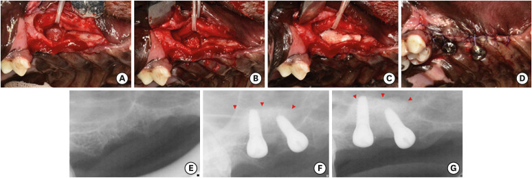
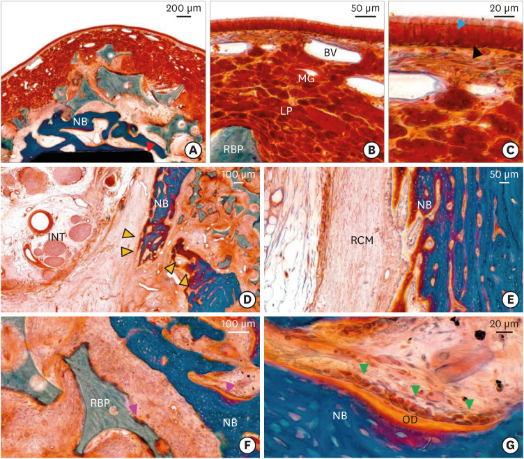
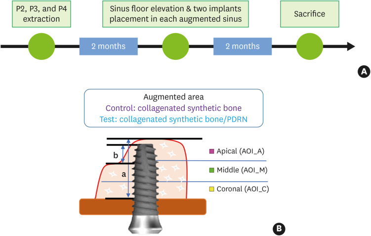
Methods: Three bimaxillary premolars (P2, P3, and P4) were extracted from 4 beagle dogs 2 months before lateral sinus floor elevation. After lateral elevation of the sinus membrane, each sinus was allocated to either the test or control group. Sinuses underwent either 1) collagenated synthetic bone graft with PDRN following lateral sinus floor elevation (test group) or 2) collagenated synthetic bone graft without PDRN after lateral sinus floor elevation (control group). Eight weeks after the surgical procedure, all animals were euthanised for a histologic and histomorphometric assessment. Augmented height (AH), protruding height (PH), and bone-to-implant contact in pristine (BICp) and augmented (BICa) bone were measured. The composition of the augmented area, which was divided into 3 areas of interest located in coronal, middle and apical areas (AOI_C, AOI_M, and AOI_A), was calculated with 3 parameters: the area percentage of new bone (pNB), residual bone graft particle (pRBP), and fibrovascular connective tissue (pFVT).
Results: AH, PH, BICp, BICa total, BICa coronal, and BICa middle values were not significantly different between sinuses in the control and test groups (all P>0.05). The BICa apical of sinuses in the test group (76.7%±9.3%) showed statistically higher values than those of sinuses in the control group (55.6%±22.1%) (P=0.038). pNB, pRBP, and pFVT showed statistically significant differences between the 2 groups in AOI_A (P=0.038, P=0.028, and P=0.007, respectively). pNB, pRBP, and pFVT in AOI_C and AOI_M were not significantly different between samples in the control and test groups (all P>0.05).
Conclusions: The histologic findings revealed that lateral sinus floor elevation with PDRN might improve early new bone formation and enable higher bone-to-implant contact.
期刊介绍:
Journal of Periodontal & Implant Science (JPIS) is a peer-reviewed and open-access journal providing up-to-date information relevant to professionalism of periodontology and dental implantology. JPIS is dedicated to global and extensive publication which includes evidence-based original articles, and fundamental reviews in order to cover a variety of interests in the field of periodontal as well as implant science.
 求助内容:
求助内容: 应助结果提醒方式:


