Cory W Baumann, Christopher P Ingalls, Dawn A Lowe
{"title":"体内偏心收缩后Mdx肌无力的机制。","authors":"Cory W Baumann, Christopher P Ingalls, Dawn A Lowe","doi":"10.1007/s10974-022-09617-1","DOIUrl":null,"url":null,"abstract":"<p><p>Skeletal muscle of the dystrophin-deficient mdx mouse is hypersensitive to eccentric (ECC) contraction-induced strength loss due to plasmalemmal electrical dysfunction. Despite plasmalemmal inexcitability being a logical mechanism responsible for weakness, it remains unclear if processes up- and/or down-stream remain functionally intact in injured mdx muscle. The purpose of this study was to analyze additional processes necessary for excitation-contraction coupling that are potentially disrupted by ECC contractions. Anterior crural muscles (tibialis anterior, extensor digitorum longus [EDL], and extensor hallucis muscles) of wildtype (WT) and mdx mice were injured in vivo with 50 ECC contractions and torque was measured immediately before and after the contraction bout. Following the in vivo assessment, EDL ex vivo isometric and caffeine forces were analyzed. In vivo isometric torque and ex vivo force in WT muscle were reduced 38 and 30% (p < 0.001), while caffeine force was also reduced (p = 0.021), albeit to a lesser degree (9%). In contrast, in vivo isometric torque, ex vivo isometric force and ex vivo caffeine-induced force were all reduced 56-67% (p < 0.001) in mdx muscle and did not differ from one another (p = 0.114). Disproportional reductions in isometric strength and caffeine-induced force confirm that ECC contractions uncoupled the plasmalemma from the ryanodine receptors (RyRs) in WT muscle. In mdx muscle, the proportional reductions in isometric strength and caffeine-induced force following ECC contractions reveal that dysfunction occurs at and/or distal to the RyRs immediately post-injury. Thus, weakness in injured mdx muscle cannot be isolated to one mechanism, rather several steps of muscle contraction are disrupted.</p>","PeriodicalId":16422,"journal":{"name":"Journal of Muscle Research and Cell Motility","volume":"43 2","pages":"63-72"},"PeriodicalIF":1.7000,"publicationDate":"2022-06-01","publicationTypes":"Journal Article","fieldsOfStudy":null,"isOpenAccess":false,"openAccessPdf":"","citationCount":"0","resultStr":"{\"title\":\"Mechanisms of weakness in Mdx muscle following in vivo eccentric contractions.\",\"authors\":\"Cory W Baumann, Christopher P Ingalls, Dawn A Lowe\",\"doi\":\"10.1007/s10974-022-09617-1\",\"DOIUrl\":null,\"url\":null,\"abstract\":\"<p><p>Skeletal muscle of the dystrophin-deficient mdx mouse is hypersensitive to eccentric (ECC) contraction-induced strength loss due to plasmalemmal electrical dysfunction. Despite plasmalemmal inexcitability being a logical mechanism responsible for weakness, it remains unclear if processes up- and/or down-stream remain functionally intact in injured mdx muscle. The purpose of this study was to analyze additional processes necessary for excitation-contraction coupling that are potentially disrupted by ECC contractions. Anterior crural muscles (tibialis anterior, extensor digitorum longus [EDL], and extensor hallucis muscles) of wildtype (WT) and mdx mice were injured in vivo with 50 ECC contractions and torque was measured immediately before and after the contraction bout. Following the in vivo assessment, EDL ex vivo isometric and caffeine forces were analyzed. In vivo isometric torque and ex vivo force in WT muscle were reduced 38 and 30% (p < 0.001), while caffeine force was also reduced (p = 0.021), albeit to a lesser degree (9%). In contrast, in vivo isometric torque, ex vivo isometric force and ex vivo caffeine-induced force were all reduced 56-67% (p < 0.001) in mdx muscle and did not differ from one another (p = 0.114). Disproportional reductions in isometric strength and caffeine-induced force confirm that ECC contractions uncoupled the plasmalemma from the ryanodine receptors (RyRs) in WT muscle. In mdx muscle, the proportional reductions in isometric strength and caffeine-induced force following ECC contractions reveal that dysfunction occurs at and/or distal to the RyRs immediately post-injury. Thus, weakness in injured mdx muscle cannot be isolated to one mechanism, rather several steps of muscle contraction are disrupted.</p>\",\"PeriodicalId\":16422,\"journal\":{\"name\":\"Journal of Muscle Research and Cell Motility\",\"volume\":\"43 2\",\"pages\":\"63-72\"},\"PeriodicalIF\":1.7000,\"publicationDate\":\"2022-06-01\",\"publicationTypes\":\"Journal Article\",\"fieldsOfStudy\":null,\"isOpenAccess\":false,\"openAccessPdf\":\"\",\"citationCount\":\"0\",\"resultStr\":null,\"platform\":\"Semanticscholar\",\"paperid\":null,\"PeriodicalName\":\"Journal of Muscle Research and Cell Motility\",\"FirstCategoryId\":\"99\",\"ListUrlMain\":\"https://doi.org/10.1007/s10974-022-09617-1\",\"RegionNum\":3,\"RegionCategory\":\"生物学\",\"ArticlePicture\":[],\"TitleCN\":null,\"AbstractTextCN\":null,\"PMCID\":null,\"EPubDate\":\"2022/4/20 0:00:00\",\"PubModel\":\"Epub\",\"JCR\":\"Q4\",\"JCRName\":\"CELL BIOLOGY\",\"Score\":null,\"Total\":0}","platform":"Semanticscholar","paperid":null,"PeriodicalName":"Journal of Muscle Research and Cell Motility","FirstCategoryId":"99","ListUrlMain":"https://doi.org/10.1007/s10974-022-09617-1","RegionNum":3,"RegionCategory":"生物学","ArticlePicture":[],"TitleCN":null,"AbstractTextCN":null,"PMCID":null,"EPubDate":"2022/4/20 0:00:00","PubModel":"Epub","JCR":"Q4","JCRName":"CELL BIOLOGY","Score":null,"Total":0}
Mechanisms of weakness in Mdx muscle following in vivo eccentric contractions.
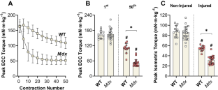
Skeletal muscle of the dystrophin-deficient mdx mouse is hypersensitive to eccentric (ECC) contraction-induced strength loss due to plasmalemmal electrical dysfunction. Despite plasmalemmal inexcitability being a logical mechanism responsible for weakness, it remains unclear if processes up- and/or down-stream remain functionally intact in injured mdx muscle. The purpose of this study was to analyze additional processes necessary for excitation-contraction coupling that are potentially disrupted by ECC contractions. Anterior crural muscles (tibialis anterior, extensor digitorum longus [EDL], and extensor hallucis muscles) of wildtype (WT) and mdx mice were injured in vivo with 50 ECC contractions and torque was measured immediately before and after the contraction bout. Following the in vivo assessment, EDL ex vivo isometric and caffeine forces were analyzed. In vivo isometric torque and ex vivo force in WT muscle were reduced 38 and 30% (p < 0.001), while caffeine force was also reduced (p = 0.021), albeit to a lesser degree (9%). In contrast, in vivo isometric torque, ex vivo isometric force and ex vivo caffeine-induced force were all reduced 56-67% (p < 0.001) in mdx muscle and did not differ from one another (p = 0.114). Disproportional reductions in isometric strength and caffeine-induced force confirm that ECC contractions uncoupled the plasmalemma from the ryanodine receptors (RyRs) in WT muscle. In mdx muscle, the proportional reductions in isometric strength and caffeine-induced force following ECC contractions reveal that dysfunction occurs at and/or distal to the RyRs immediately post-injury. Thus, weakness in injured mdx muscle cannot be isolated to one mechanism, rather several steps of muscle contraction are disrupted.
期刊介绍:
The Journal of Muscle Research and Cell Motility has as its main aim the publication of original research which bears on either the excitation and contraction of muscle, the analysis of any one of the processes involved therein, the processes underlying contractility and motility of animal and plant cells, the toxicology and pharmacology related to contractility, or the formation, dynamics and turnover of contractile structures in muscle and non-muscle cells. Studies describing the impact of pathogenic mutations in genes encoding components of contractile structures in humans or animals are welcome, provided they offer mechanistic insight into the disease process or the underlying gene function. The policy of the Journal is to encourage any form of novel practical study whatever its specialist interest, as long as it falls within this broad field. Theoretical essays are welcome provided that they are concise and suggest practical ways in which they may be tested. Manuscripts reporting new mutations in known disease genes without validation and mechanistic insight will not be considered. It is the policy of the journal that cells lines, hybridomas and DNA clones should be made available by the developers to any qualified investigator. Submission of a manuscript for publication constitutes an agreement of the authors to abide by this principle.
 求助内容:
求助内容: 应助结果提醒方式:


