Akos Varga-Szemes, Pal Maurovich-Horvat, U Joseph Schoepf, Emese Zsarnoczay, Robert Pelberg, Gregg W Stone, Matthew J Budoff
{"title":"冠状动脉粥样硬化的计算机断层扫描评估:从基于阈值的评估到组织学验证的斑块定量。","authors":"Akos Varga-Szemes, Pal Maurovich-Horvat, U Joseph Schoepf, Emese Zsarnoczay, Robert Pelberg, Gregg W Stone, Matthew J Budoff","doi":"10.1097/RTI.0000000000000711","DOIUrl":null,"url":null,"abstract":"<p><p>Arterial plaque rupture and thrombosis is the primary cause of major cardiovascular and neurovascular events. The identification of atherosclerosis, especially high-risk plaques, is therefore crucial to identify high-risk patients and to implement preventive therapies. Computed tomography angiography has the ability to visualize and characterize vascular plaques. The standard methods for plaque evaluation rely on the assessment of plaque burden, stenosis severity, the presence of positive remodeling, napkin ring sign, and spotty calcification, as well as Hounsfield Unit (HU)-based thresholding for plaque quantification; the latter with multiple shortcomings. Semiautomated threshold-based segmentation techniques with predefined HU ranges identify and quantify limited plaque characteristics, such as low attenuation, non-calcified, and calcified plaque components. Contrary to HU-based thresholds, histologically validated plaque characterization, and quantification, an emerging Artificial intelligence-based approach has the ability to differentiate specific tissue types based on a biological correlate, such as lipid-rich necrotic core and intraplaque hemorrhage that determine plaque vulnerability. In this article, we review the relevance of plaque characterization and quantification and discuss the benefits and limitations of the currently available plaque assessment and classification techniques.</p>","PeriodicalId":49974,"journal":{"name":"Journal of Thoracic Imaging","volume":null,"pages":null},"PeriodicalIF":2.0000,"publicationDate":"2023-07-01","publicationTypes":"Journal Article","fieldsOfStudy":null,"isOpenAccess":false,"openAccessPdf":"https://ftp.ncbi.nlm.nih.gov/pub/pmc/oa_pdf/46/3b/rti-38-226.PMC10287054.pdf","citationCount":"2","resultStr":"{\"title\":\"Computed Tomography Assessment of Coronary Atherosclerosis: From Threshold-Based Evaluation to Histologically Validated Plaque Quantification.\",\"authors\":\"Akos Varga-Szemes, Pal Maurovich-Horvat, U Joseph Schoepf, Emese Zsarnoczay, Robert Pelberg, Gregg W Stone, Matthew J Budoff\",\"doi\":\"10.1097/RTI.0000000000000711\",\"DOIUrl\":null,\"url\":null,\"abstract\":\"<p><p>Arterial plaque rupture and thrombosis is the primary cause of major cardiovascular and neurovascular events. The identification of atherosclerosis, especially high-risk plaques, is therefore crucial to identify high-risk patients and to implement preventive therapies. Computed tomography angiography has the ability to visualize and characterize vascular plaques. The standard methods for plaque evaluation rely on the assessment of plaque burden, stenosis severity, the presence of positive remodeling, napkin ring sign, and spotty calcification, as well as Hounsfield Unit (HU)-based thresholding for plaque quantification; the latter with multiple shortcomings. Semiautomated threshold-based segmentation techniques with predefined HU ranges identify and quantify limited plaque characteristics, such as low attenuation, non-calcified, and calcified plaque components. Contrary to HU-based thresholds, histologically validated plaque characterization, and quantification, an emerging Artificial intelligence-based approach has the ability to differentiate specific tissue types based on a biological correlate, such as lipid-rich necrotic core and intraplaque hemorrhage that determine plaque vulnerability. In this article, we review the relevance of plaque characterization and quantification and discuss the benefits and limitations of the currently available plaque assessment and classification techniques.</p>\",\"PeriodicalId\":49974,\"journal\":{\"name\":\"Journal of Thoracic Imaging\",\"volume\":null,\"pages\":null},\"PeriodicalIF\":2.0000,\"publicationDate\":\"2023-07-01\",\"publicationTypes\":\"Journal Article\",\"fieldsOfStudy\":null,\"isOpenAccess\":false,\"openAccessPdf\":\"https://ftp.ncbi.nlm.nih.gov/pub/pmc/oa_pdf/46/3b/rti-38-226.PMC10287054.pdf\",\"citationCount\":\"2\",\"resultStr\":null,\"platform\":\"Semanticscholar\",\"paperid\":null,\"PeriodicalName\":\"Journal of Thoracic Imaging\",\"FirstCategoryId\":\"3\",\"ListUrlMain\":\"https://doi.org/10.1097/RTI.0000000000000711\",\"RegionNum\":4,\"RegionCategory\":\"医学\",\"ArticlePicture\":[],\"TitleCN\":null,\"AbstractTextCN\":null,\"PMCID\":null,\"EPubDate\":\"2023/4/10 0:00:00\",\"PubModel\":\"Epub\",\"JCR\":\"Q3\",\"JCRName\":\"RADIOLOGY, NUCLEAR MEDICINE & MEDICAL IMAGING\",\"Score\":null,\"Total\":0}","platform":"Semanticscholar","paperid":null,"PeriodicalName":"Journal of Thoracic Imaging","FirstCategoryId":"3","ListUrlMain":"https://doi.org/10.1097/RTI.0000000000000711","RegionNum":4,"RegionCategory":"医学","ArticlePicture":[],"TitleCN":null,"AbstractTextCN":null,"PMCID":null,"EPubDate":"2023/4/10 0:00:00","PubModel":"Epub","JCR":"Q3","JCRName":"RADIOLOGY, NUCLEAR MEDICINE & MEDICAL IMAGING","Score":null,"Total":0}
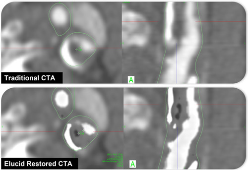
Computed Tomography Assessment of Coronary Atherosclerosis: From Threshold-Based Evaluation to Histologically Validated Plaque Quantification.
Arterial plaque rupture and thrombosis is the primary cause of major cardiovascular and neurovascular events. The identification of atherosclerosis, especially high-risk plaques, is therefore crucial to identify high-risk patients and to implement preventive therapies. Computed tomography angiography has the ability to visualize and characterize vascular plaques. The standard methods for plaque evaluation rely on the assessment of plaque burden, stenosis severity, the presence of positive remodeling, napkin ring sign, and spotty calcification, as well as Hounsfield Unit (HU)-based thresholding for plaque quantification; the latter with multiple shortcomings. Semiautomated threshold-based segmentation techniques with predefined HU ranges identify and quantify limited plaque characteristics, such as low attenuation, non-calcified, and calcified plaque components. Contrary to HU-based thresholds, histologically validated plaque characterization, and quantification, an emerging Artificial intelligence-based approach has the ability to differentiate specific tissue types based on a biological correlate, such as lipid-rich necrotic core and intraplaque hemorrhage that determine plaque vulnerability. In this article, we review the relevance of plaque characterization and quantification and discuss the benefits and limitations of the currently available plaque assessment and classification techniques.
期刊介绍:
Journal of Thoracic Imaging (JTI) provides authoritative information on all aspects of the use of imaging techniques in the diagnosis of cardiac and pulmonary diseases. Original articles and analytical reviews published in this timely journal provide the very latest thinking of leading experts concerning the use of chest radiography, computed tomography, magnetic resonance imaging, positron emission tomography, ultrasound, and all other promising imaging techniques in cardiopulmonary radiology.
Official Journal of the Society of Thoracic Radiology:
Japanese Society of Thoracic Radiology
Korean Society of Thoracic Radiology
European Society of Thoracic Imaging.


 求助内容:
求助内容: 应助结果提醒方式:


