Eman A B Aguori, Nilüfer Ersan, Zehra S Dölekoğlu, Dilhan Ilgüy
{"title":"健康后牙靠近上颌窦底与粘膜增厚的关系:一项CBCT研究","authors":"Eman A B Aguori, Nilüfer Ersan, Zehra S Dölekoğlu, Dilhan Ilgüy","doi":"10.1007/s11282-022-00666-3","DOIUrl":null,"url":null,"abstract":"<p><strong>Objectives: </strong>To evaluate the relationship between proximity of the root apices of healthy maxillary posterior teeth to the maxillary sinus floor (MSF) and mucosal thickening (MT) of the MSF using cone beam computed tomography (CBCT).</p><p><strong>Methods: </strong>Eighty-four CBCT images obtained from the patients, aged between 20 and 70 years with healthy and fully dentate maxillary posterior sextants, were included. The anatomical relationship between root apices of maxillary posterior teeth and MSF, was evaluated: (Type 1: no contact, Type 2: at least one root apex in contact, Type 3: at least one root apex protruding into MSF). Besides, MT of the MSF was measured from the thickest region. The patients were categorized into two groups based on the absence (≤ 2 mm) or the presence (2 < mm) of MT. Statistical significance was accepted at p < 0.05.</p><p><strong>Results: </strong>Intraexaminer consistency demonstrated an excellent agreement (p < 0.05). The prevalence of Type 1, 2, and 3 proximity were found as 26 (15.5%), 61 (36.3%), and 81 (48.2%); respectively. Overall, 62 (36.9%) maxillary sinuses demonstrated MT (2 < mm, mean: 8.6 ± 7.5 mm). The prevalence and mean values of MT (2 < mm) were not found to be statistically significantly different in terms of sex and proximity types (p > 0.05). Logistic regression analysis results were not found to be statistically significant (p > 0.05).</p><p><strong>Conclusion: </strong>The proximity of healthy maxillary posterior teeth to the MSF was not found to be a contributing factor for the MT of the MSF. Further studies with larger samples, taking the other factors causing MT into consideration, are needed.</p>","PeriodicalId":56103,"journal":{"name":"Oral Radiology","volume":null,"pages":null},"PeriodicalIF":1.6000,"publicationDate":"2023-07-01","publicationTypes":"Journal Article","fieldsOfStudy":null,"isOpenAccess":false,"openAccessPdf":"","citationCount":"0","resultStr":"{\"title\":\"Proximity of healthy posterior teeth to the maxillary sinus floor in relation to mucosal thickening: a CBCT study.\",\"authors\":\"Eman A B Aguori, Nilüfer Ersan, Zehra S Dölekoğlu, Dilhan Ilgüy\",\"doi\":\"10.1007/s11282-022-00666-3\",\"DOIUrl\":null,\"url\":null,\"abstract\":\"<p><strong>Objectives: </strong>To evaluate the relationship between proximity of the root apices of healthy maxillary posterior teeth to the maxillary sinus floor (MSF) and mucosal thickening (MT) of the MSF using cone beam computed tomography (CBCT).</p><p><strong>Methods: </strong>Eighty-four CBCT images obtained from the patients, aged between 20 and 70 years with healthy and fully dentate maxillary posterior sextants, were included. The anatomical relationship between root apices of maxillary posterior teeth and MSF, was evaluated: (Type 1: no contact, Type 2: at least one root apex in contact, Type 3: at least one root apex protruding into MSF). Besides, MT of the MSF was measured from the thickest region. The patients were categorized into two groups based on the absence (≤ 2 mm) or the presence (2 < mm) of MT. Statistical significance was accepted at p < 0.05.</p><p><strong>Results: </strong>Intraexaminer consistency demonstrated an excellent agreement (p < 0.05). The prevalence of Type 1, 2, and 3 proximity were found as 26 (15.5%), 61 (36.3%), and 81 (48.2%); respectively. Overall, 62 (36.9%) maxillary sinuses demonstrated MT (2 < mm, mean: 8.6 ± 7.5 mm). The prevalence and mean values of MT (2 < mm) were not found to be statistically significantly different in terms of sex and proximity types (p > 0.05). Logistic regression analysis results were not found to be statistically significant (p > 0.05).</p><p><strong>Conclusion: </strong>The proximity of healthy maxillary posterior teeth to the MSF was not found to be a contributing factor for the MT of the MSF. Further studies with larger samples, taking the other factors causing MT into consideration, are needed.</p>\",\"PeriodicalId\":56103,\"journal\":{\"name\":\"Oral Radiology\",\"volume\":null,\"pages\":null},\"PeriodicalIF\":1.6000,\"publicationDate\":\"2023-07-01\",\"publicationTypes\":\"Journal Article\",\"fieldsOfStudy\":null,\"isOpenAccess\":false,\"openAccessPdf\":\"\",\"citationCount\":\"0\",\"resultStr\":null,\"platform\":\"Semanticscholar\",\"paperid\":null,\"PeriodicalName\":\"Oral Radiology\",\"FirstCategoryId\":\"3\",\"ListUrlMain\":\"https://doi.org/10.1007/s11282-022-00666-3\",\"RegionNum\":3,\"RegionCategory\":\"医学\",\"ArticlePicture\":[],\"TitleCN\":null,\"AbstractTextCN\":null,\"PMCID\":null,\"EPubDate\":\"\",\"PubModel\":\"\",\"JCR\":\"Q3\",\"JCRName\":\"DENTISTRY, ORAL SURGERY & MEDICINE\",\"Score\":null,\"Total\":0}","platform":"Semanticscholar","paperid":null,"PeriodicalName":"Oral Radiology","FirstCategoryId":"3","ListUrlMain":"https://doi.org/10.1007/s11282-022-00666-3","RegionNum":3,"RegionCategory":"医学","ArticlePicture":[],"TitleCN":null,"AbstractTextCN":null,"PMCID":null,"EPubDate":"","PubModel":"","JCR":"Q3","JCRName":"DENTISTRY, ORAL SURGERY & MEDICINE","Score":null,"Total":0}
Proximity of healthy posterior teeth to the maxillary sinus floor in relation to mucosal thickening: a CBCT study.
Objectives: To evaluate the relationship between proximity of the root apices of healthy maxillary posterior teeth to the maxillary sinus floor (MSF) and mucosal thickening (MT) of the MSF using cone beam computed tomography (CBCT).
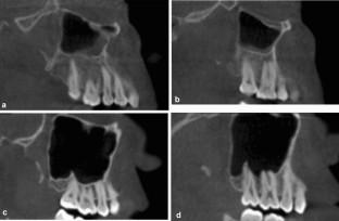
Methods: Eighty-four CBCT images obtained from the patients, aged between 20 and 70 years with healthy and fully dentate maxillary posterior sextants, were included. The anatomical relationship between root apices of maxillary posterior teeth and MSF, was evaluated: (Type 1: no contact, Type 2: at least one root apex in contact, Type 3: at least one root apex protruding into MSF). Besides, MT of the MSF was measured from the thickest region. The patients were categorized into two groups based on the absence (≤ 2 mm) or the presence (2 < mm) of MT. Statistical significance was accepted at p < 0.05.
Results: Intraexaminer consistency demonstrated an excellent agreement (p < 0.05). The prevalence of Type 1, 2, and 3 proximity were found as 26 (15.5%), 61 (36.3%), and 81 (48.2%); respectively. Overall, 62 (36.9%) maxillary sinuses demonstrated MT (2 < mm, mean: 8.6 ± 7.5 mm). The prevalence and mean values of MT (2 < mm) were not found to be statistically significantly different in terms of sex and proximity types (p > 0.05). Logistic regression analysis results were not found to be statistically significant (p > 0.05).
Conclusion: The proximity of healthy maxillary posterior teeth to the MSF was not found to be a contributing factor for the MT of the MSF. Further studies with larger samples, taking the other factors causing MT into consideration, are needed.
期刊介绍:
As the official English-language journal of the Japanese Society for Oral and Maxillofacial Radiology and the Asian Academy of Oral and Maxillofacial Radiology, Oral Radiology is intended to be a forum for international collaboration in head and neck diagnostic imaging and all related fields. Oral Radiology features cutting-edge research papers, review articles, case reports, and technical notes from both the clinical and experimental fields. As membership in the Society is not a prerequisite, contributions are welcome from researchers and clinicians worldwide.

 求助内容:
求助内容: 应助结果提醒方式:


