Fatma Öz Atalay, Fatma Gündoğdu, Gözde Elif Taşar Kapaklı, Ali Can Güneş, Yeşim Gaye Güler, Alp Usubütün
{"title":"中间滋养细胞的妊娠滋养细胞瘤:上皮样滋养细胞瘤和胎盘部位滋养细胞瘤,形态学、免疫组织化学和下一代测序的研究。","authors":"Fatma Öz Atalay, Fatma Gündoğdu, Gözde Elif Taşar Kapaklı, Ali Can Güneş, Yeşim Gaye Güler, Alp Usubütün","doi":"10.4274/tjod.galenos.2023.73549","DOIUrl":null,"url":null,"abstract":"<p><strong>Objective: </strong>Gestational trophoblastic tumors are very rare neoplasms. We determined the distinctive morphological, immunohistochemical, and clinical features of placental site trophoblastic tumors (PSTT) and epithelioid trophoblastic tumors (ETT) in our cohort.</p><p><strong>Materials and methods: </strong>Nine cases of PSTT and four cases of ETT were retrieved from the archives. Histomorphologic, immunohistochemical, and clinical features were noted. A molecular study was performed on one PSTT and one ETT case using next-generation sequencing.</p><p><strong>Results: </strong>While the nodular pattern, geographic necrosis, and extracellular eosinophilic globules were peculiar to ETTs, vessel wall affinity, marked pleomorphism, intranuclear pseudoinclusion, spindle tumor cell, and vacuolar degeneration were more specific for PSTTs in our series. An immunohistochemical panel of p63, hPL, and CD146 were helpful for the exact typing of the tumor. p63 positivity supports the ETT and diffuse staining of hPL and CD146 supports the PSTT diagnosis. Three of the patients with metastatic disease (lung and brain metastasis) except one have a high mitotic count (12 and 8) and a long interval between (8 and 10 years) antecedent pregnancy and diagnosis. While KIT and TP53 mutations were observed only in PSTT, amino acid changes in KDR, APC, and SMAD4 genes were detected both in the ETT and PSTT cases.</p><p><strong>Conclusion: </strong>In the prediction of metastasis, the long intervals between antecedent pregnancy and diagnosis, deep myometrial invasion, mitotic count, and Ki67 proliferation index were involved rather than other histomorphological parameters, but none of the parameters is an absolute predictor of the metastasis.</p>","PeriodicalId":45340,"journal":{"name":"Turkish Journal of Obstetrics and Gynecology","volume":"20 2","pages":"105-112"},"PeriodicalIF":1.3000,"publicationDate":"2023-06-01","publicationTypes":"Journal Article","fieldsOfStudy":null,"isOpenAccess":false,"openAccessPdf":"https://ftp.ncbi.nlm.nih.gov/pub/pmc/oa_pdf/d8/57/TJOG-20-105.PMC10236228.pdf","citationCount":"0","resultStr":"{\"title\":\"Gestational trophoblastic neoplasia of intermediate trophoblasts: Epithelioid trophoblastic tumor and placental site trophoblastic tumor, a study of morphologic, immunohistochemical, and next generation sequencing.\",\"authors\":\"Fatma Öz Atalay, Fatma Gündoğdu, Gözde Elif Taşar Kapaklı, Ali Can Güneş, Yeşim Gaye Güler, Alp Usubütün\",\"doi\":\"10.4274/tjod.galenos.2023.73549\",\"DOIUrl\":null,\"url\":null,\"abstract\":\"<p><strong>Objective: </strong>Gestational trophoblastic tumors are very rare neoplasms. We determined the distinctive morphological, immunohistochemical, and clinical features of placental site trophoblastic tumors (PSTT) and epithelioid trophoblastic tumors (ETT) in our cohort.</p><p><strong>Materials and methods: </strong>Nine cases of PSTT and four cases of ETT were retrieved from the archives. Histomorphologic, immunohistochemical, and clinical features were noted. A molecular study was performed on one PSTT and one ETT case using next-generation sequencing.</p><p><strong>Results: </strong>While the nodular pattern, geographic necrosis, and extracellular eosinophilic globules were peculiar to ETTs, vessel wall affinity, marked pleomorphism, intranuclear pseudoinclusion, spindle tumor cell, and vacuolar degeneration were more specific for PSTTs in our series. An immunohistochemical panel of p63, hPL, and CD146 were helpful for the exact typing of the tumor. p63 positivity supports the ETT and diffuse staining of hPL and CD146 supports the PSTT diagnosis. Three of the patients with metastatic disease (lung and brain metastasis) except one have a high mitotic count (12 and 8) and a long interval between (8 and 10 years) antecedent pregnancy and diagnosis. While KIT and TP53 mutations were observed only in PSTT, amino acid changes in KDR, APC, and SMAD4 genes were detected both in the ETT and PSTT cases.</p><p><strong>Conclusion: </strong>In the prediction of metastasis, the long intervals between antecedent pregnancy and diagnosis, deep myometrial invasion, mitotic count, and Ki67 proliferation index were involved rather than other histomorphological parameters, but none of the parameters is an absolute predictor of the metastasis.</p>\",\"PeriodicalId\":45340,\"journal\":{\"name\":\"Turkish Journal of Obstetrics and Gynecology\",\"volume\":\"20 2\",\"pages\":\"105-112\"},\"PeriodicalIF\":1.3000,\"publicationDate\":\"2023-06-01\",\"publicationTypes\":\"Journal Article\",\"fieldsOfStudy\":null,\"isOpenAccess\":false,\"openAccessPdf\":\"https://ftp.ncbi.nlm.nih.gov/pub/pmc/oa_pdf/d8/57/TJOG-20-105.PMC10236228.pdf\",\"citationCount\":\"0\",\"resultStr\":null,\"platform\":\"Semanticscholar\",\"paperid\":null,\"PeriodicalName\":\"Turkish Journal of Obstetrics and Gynecology\",\"FirstCategoryId\":\"1085\",\"ListUrlMain\":\"https://doi.org/10.4274/tjod.galenos.2023.73549\",\"RegionNum\":0,\"RegionCategory\":null,\"ArticlePicture\":[],\"TitleCN\":null,\"AbstractTextCN\":null,\"PMCID\":null,\"EPubDate\":\"\",\"PubModel\":\"\",\"JCR\":\"Q4\",\"JCRName\":\"OBSTETRICS & GYNECOLOGY\",\"Score\":null,\"Total\":0}","platform":"Semanticscholar","paperid":null,"PeriodicalName":"Turkish Journal of Obstetrics and Gynecology","FirstCategoryId":"1085","ListUrlMain":"https://doi.org/10.4274/tjod.galenos.2023.73549","RegionNum":0,"RegionCategory":null,"ArticlePicture":[],"TitleCN":null,"AbstractTextCN":null,"PMCID":null,"EPubDate":"","PubModel":"","JCR":"Q4","JCRName":"OBSTETRICS & GYNECOLOGY","Score":null,"Total":0}
Gestational trophoblastic neoplasia of intermediate trophoblasts: Epithelioid trophoblastic tumor and placental site trophoblastic tumor, a study of morphologic, immunohistochemical, and next generation sequencing.
Objective: Gestational trophoblastic tumors are very rare neoplasms. We determined the distinctive morphological, immunohistochemical, and clinical features of placental site trophoblastic tumors (PSTT) and epithelioid trophoblastic tumors (ETT) in our cohort.
Materials and methods: Nine cases of PSTT and four cases of ETT were retrieved from the archives. Histomorphologic, immunohistochemical, and clinical features were noted. A molecular study was performed on one PSTT and one ETT case using next-generation sequencing.
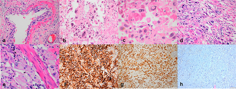
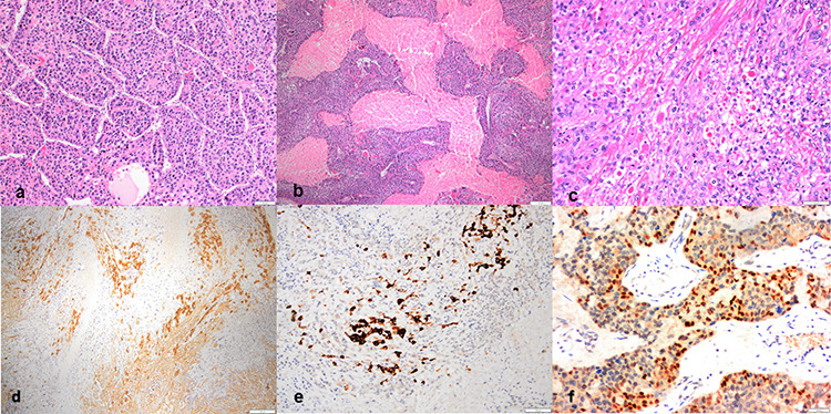
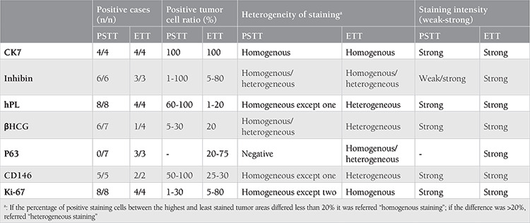
Results: While the nodular pattern, geographic necrosis, and extracellular eosinophilic globules were peculiar to ETTs, vessel wall affinity, marked pleomorphism, intranuclear pseudoinclusion, spindle tumor cell, and vacuolar degeneration were more specific for PSTTs in our series. An immunohistochemical panel of p63, hPL, and CD146 were helpful for the exact typing of the tumor. p63 positivity supports the ETT and diffuse staining of hPL and CD146 supports the PSTT diagnosis. Three of the patients with metastatic disease (lung and brain metastasis) except one have a high mitotic count (12 and 8) and a long interval between (8 and 10 years) antecedent pregnancy and diagnosis. While KIT and TP53 mutations were observed only in PSTT, amino acid changes in KDR, APC, and SMAD4 genes were detected both in the ETT and PSTT cases.
Conclusion: In the prediction of metastasis, the long intervals between antecedent pregnancy and diagnosis, deep myometrial invasion, mitotic count, and Ki67 proliferation index were involved rather than other histomorphological parameters, but none of the parameters is an absolute predictor of the metastasis.

 求助内容:
求助内容: 应助结果提醒方式:


