In Gul Kim, Yanru Wu, Su A Park, Ji Suk Choi, Seong Keun Kwon, Seung Hong Choi, Kyeong Cheon Jung, Jung-Woog Shin, Eun-Jae Chung
{"title":"通过生物反应器培养合成支架在犬模型中重建食管的评估。","authors":"In Gul Kim, Yanru Wu, Su A Park, Ji Suk Choi, Seong Keun Kwon, Seung Hong Choi, Kyeong Cheon Jung, Jung-Woog Shin, Eun-Jae Chung","doi":"10.21053/ceo.2022.01522","DOIUrl":null,"url":null,"abstract":"<p><strong>Objectives: </strong>Using tissue-engineered materials for esophageal reconstruction is a technically challenging task in animals that requires bioreactor training to enhance cellular reactivity. There have been many attempts at esophageal tissue engineering, but the success rate has been limited due to difficulty in initial epithelialization in the special environment of peristalsis. The purpose of this study was to evaluate the potential of an artificial esophagus that can enhance the regeneration of esophageal mucosa and muscle through the optimal combination of a double-layered polymeric scaffold and a custom-designed mesenchymal stem cell-based bioreactor system in a canine model.</p><p><strong>Methods: </strong>We fabricated a novel double-layered scaffold as a tissue-engineered esophagus using an electrospinning technique. Prior to transplantation, human-derived mesenchymal stem cells were seeded into the lumen of the scaffold, and bioreactor cultivation was performed to enhance cellular reactivity. After 3 days of cultivation using the bioreactor system, tissue-engineered artificial esophagus was transplanted into a partial esophageal defect (5×3 cm-long resection) in a canine model.</p><p><strong>Results: </strong>Scanning electron microscopy (SEM) showed that the electrospun fibers in a tubular scaffold were randomly and circumferentially located toward the inner and outer surfaces. Complete recovery of the esophageal mucosa was confirmed by endoscopic analysis and SEM. Esophagogastroduodenoscopy and computed tomography also showed that there were no signs of leakage or stricture and that there was a normal lumen with complete epithelialization. Significant regeneration of the mucosal layer was observed by keratin-5 immunostaining. Alpha-smooth muscle actin immunostaining showed significantly greater esophageal muscle regeneration at 12 months than at 6 months.</p><p><strong>Conclusion: </strong>Custom-designed bioreactor cultured electrospun polyurethane scaffolds can be a promising approach for esophageal tissue engineering.</p>","PeriodicalId":10318,"journal":{"name":"Clinical and Experimental Otorhinolaryngology","volume":"16 2","pages":"165-176"},"PeriodicalIF":2.9000,"publicationDate":"2023-05-01","publicationTypes":"Journal Article","fieldsOfStudy":null,"isOpenAccess":false,"openAccessPdf":"https://ftp.ncbi.nlm.nih.gov/pub/pmc/oa_pdf/f5/25/ceo-2022-01522.PMC10208848.pdf","citationCount":"1","resultStr":"{\"title\":\"Assessment of Esophageal Reconstruction via Bioreactor Cultivation of a Synthetic Scaffold in a Canine Model.\",\"authors\":\"In Gul Kim, Yanru Wu, Su A Park, Ji Suk Choi, Seong Keun Kwon, Seung Hong Choi, Kyeong Cheon Jung, Jung-Woog Shin, Eun-Jae Chung\",\"doi\":\"10.21053/ceo.2022.01522\",\"DOIUrl\":null,\"url\":null,\"abstract\":\"<p><strong>Objectives: </strong>Using tissue-engineered materials for esophageal reconstruction is a technically challenging task in animals that requires bioreactor training to enhance cellular reactivity. There have been many attempts at esophageal tissue engineering, but the success rate has been limited due to difficulty in initial epithelialization in the special environment of peristalsis. The purpose of this study was to evaluate the potential of an artificial esophagus that can enhance the regeneration of esophageal mucosa and muscle through the optimal combination of a double-layered polymeric scaffold and a custom-designed mesenchymal stem cell-based bioreactor system in a canine model.</p><p><strong>Methods: </strong>We fabricated a novel double-layered scaffold as a tissue-engineered esophagus using an electrospinning technique. Prior to transplantation, human-derived mesenchymal stem cells were seeded into the lumen of the scaffold, and bioreactor cultivation was performed to enhance cellular reactivity. After 3 days of cultivation using the bioreactor system, tissue-engineered artificial esophagus was transplanted into a partial esophageal defect (5×3 cm-long resection) in a canine model.</p><p><strong>Results: </strong>Scanning electron microscopy (SEM) showed that the electrospun fibers in a tubular scaffold were randomly and circumferentially located toward the inner and outer surfaces. Complete recovery of the esophageal mucosa was confirmed by endoscopic analysis and SEM. Esophagogastroduodenoscopy and computed tomography also showed that there were no signs of leakage or stricture and that there was a normal lumen with complete epithelialization. Significant regeneration of the mucosal layer was observed by keratin-5 immunostaining. Alpha-smooth muscle actin immunostaining showed significantly greater esophageal muscle regeneration at 12 months than at 6 months.</p><p><strong>Conclusion: </strong>Custom-designed bioreactor cultured electrospun polyurethane scaffolds can be a promising approach for esophageal tissue engineering.</p>\",\"PeriodicalId\":10318,\"journal\":{\"name\":\"Clinical and Experimental Otorhinolaryngology\",\"volume\":\"16 2\",\"pages\":\"165-176\"},\"PeriodicalIF\":2.9000,\"publicationDate\":\"2023-05-01\",\"publicationTypes\":\"Journal Article\",\"fieldsOfStudy\":null,\"isOpenAccess\":false,\"openAccessPdf\":\"https://ftp.ncbi.nlm.nih.gov/pub/pmc/oa_pdf/f5/25/ceo-2022-01522.PMC10208848.pdf\",\"citationCount\":\"1\",\"resultStr\":null,\"platform\":\"Semanticscholar\",\"paperid\":null,\"PeriodicalName\":\"Clinical and Experimental Otorhinolaryngology\",\"FirstCategoryId\":\"3\",\"ListUrlMain\":\"https://doi.org/10.21053/ceo.2022.01522\",\"RegionNum\":3,\"RegionCategory\":\"医学\",\"ArticlePicture\":[],\"TitleCN\":null,\"AbstractTextCN\":null,\"PMCID\":null,\"EPubDate\":\"\",\"PubModel\":\"\",\"JCR\":\"Q1\",\"JCRName\":\"OTORHINOLARYNGOLOGY\",\"Score\":null,\"Total\":0}","platform":"Semanticscholar","paperid":null,"PeriodicalName":"Clinical and Experimental Otorhinolaryngology","FirstCategoryId":"3","ListUrlMain":"https://doi.org/10.21053/ceo.2022.01522","RegionNum":3,"RegionCategory":"医学","ArticlePicture":[],"TitleCN":null,"AbstractTextCN":null,"PMCID":null,"EPubDate":"","PubModel":"","JCR":"Q1","JCRName":"OTORHINOLARYNGOLOGY","Score":null,"Total":0}
Assessment of Esophageal Reconstruction via Bioreactor Cultivation of a Synthetic Scaffold in a Canine Model.
Objectives: Using tissue-engineered materials for esophageal reconstruction is a technically challenging task in animals that requires bioreactor training to enhance cellular reactivity. There have been many attempts at esophageal tissue engineering, but the success rate has been limited due to difficulty in initial epithelialization in the special environment of peristalsis. The purpose of this study was to evaluate the potential of an artificial esophagus that can enhance the regeneration of esophageal mucosa and muscle through the optimal combination of a double-layered polymeric scaffold and a custom-designed mesenchymal stem cell-based bioreactor system in a canine model.
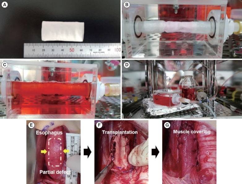
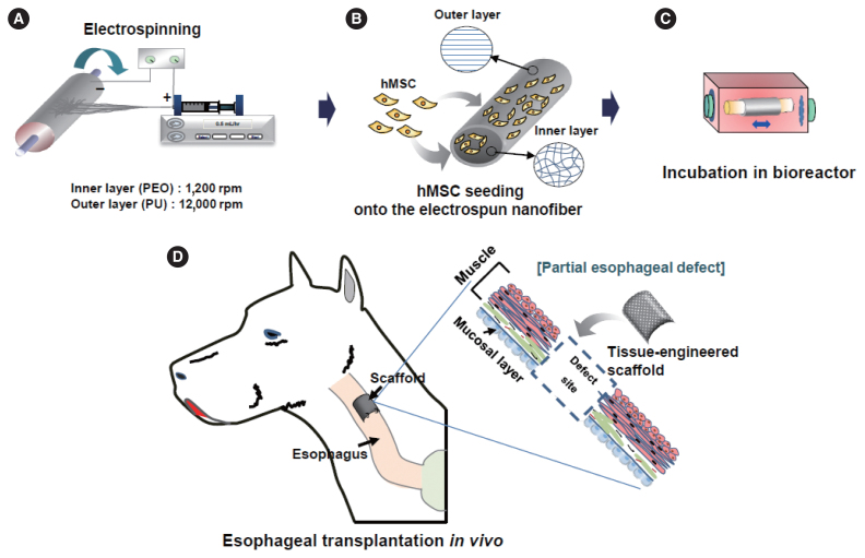
Methods: We fabricated a novel double-layered scaffold as a tissue-engineered esophagus using an electrospinning technique. Prior to transplantation, human-derived mesenchymal stem cells were seeded into the lumen of the scaffold, and bioreactor cultivation was performed to enhance cellular reactivity. After 3 days of cultivation using the bioreactor system, tissue-engineered artificial esophagus was transplanted into a partial esophageal defect (5×3 cm-long resection) in a canine model.
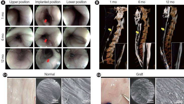
Results: Scanning electron microscopy (SEM) showed that the electrospun fibers in a tubular scaffold were randomly and circumferentially located toward the inner and outer surfaces. Complete recovery of the esophageal mucosa was confirmed by endoscopic analysis and SEM. Esophagogastroduodenoscopy and computed tomography also showed that there were no signs of leakage or stricture and that there was a normal lumen with complete epithelialization. Significant regeneration of the mucosal layer was observed by keratin-5 immunostaining. Alpha-smooth muscle actin immunostaining showed significantly greater esophageal muscle regeneration at 12 months than at 6 months.
Conclusion: Custom-designed bioreactor cultured electrospun polyurethane scaffolds can be a promising approach for esophageal tissue engineering.
期刊介绍:
Clinical and Experimental Otorhinolaryngology (Clin Exp Otorhinolaryngol, CEO) is an international peer-reviewed journal on recent developments in diagnosis and treatment of otorhinolaryngology-head and neck surgery and dedicated to the advancement of patient care in ear, nose, throat, head, and neck disorders. This journal publishes original articles relating to both clinical and basic researches, reviews, and clinical trials, encompassing the whole topics of otorhinolaryngology-head and neck surgery.
CEO was first issued in 2008 and this journal is published in English four times (the last day of February, May, August, and November) per year by the Korean Society of Otorhinolaryngology-Head and Neck Surgery. The Journal aims at publishing evidence-based, scientifically written articles from different disciplines of otorhinolaryngology field.
The readership contains clinical/basic research into current practice in otorhinolaryngology, audiology, speech pathology, head and neck oncology, plastic and reconstructive surgery. The readers are otolaryngologists, head and neck surgeons and oncologists, audiologists, and speech pathologists.
 求助内容:
求助内容: 应助结果提醒方式:


