Dmitrii S Maltsev, Alexei N Kulikov, Maria A Burnasheva
{"title":"用光学相干断层血管造影记录高眼压患者的脉动性眼血流。","authors":"Dmitrii S Maltsev, Alexei N Kulikov, Maria A Burnasheva","doi":"10.4103/joco.joco_161_22","DOIUrl":null,"url":null,"abstract":"<p><strong>Purpose: </strong>To present a series of cases demonstrating pulsatile ocular blood flow registered with optical coherence tomography angiography (OCTA) and to describe the clinical characteristics of this phenomenon.</p><p><strong>Methods: </strong>Seven primary open-angle glaucoma patients (eight eyes) were included, with a median age of 67.0 years (range, 39-73 years), who demonstrated alternating hypointense bands of OCTA flow signal on the macular scan at increased intraocular pressure (IOP). All patients received comprehensive ophthalmic examination, OCTA examination with RTVue-XR, and infrared video scanning laser ophthalmoscopy. Changes in retinal microcirculation were assessed on the raw OCTA scans as well as the resultant vessel density maps before and after IOP reduction.</p><p><strong>Results: </strong>Median IOP in study eyes was 39.0 mmHg (range, 36-58 mmHg). Hypointense bands of OCTA flow signal were associated with arterial pulsation on video scanning laser ophthalmoscopy in all eyes and agreed with the heart rate and resulted in a spotted grid pattern of hypoperfusion on vessel density maps in seven eyes. Median vessel density in superficial capillary plexus and deep capillary plexus was 32.4% and 47.2%, respectively, at high IOP, and increased statistically significantly to 36.5% (<i>P</i> = 0.016) and 50.9% (<i>P</i> = 0.016), respectively, after IOP reduction.</p><p><strong>Conclusions: </strong>Alternating hypointense flow signal bands on OCTA scans are possibly caused by the pulsatile character of retinal blood flow during the cardiac cycle in eyes with high IOP and may reflect the imbalance between IOP and perfusion pressure. This phenomenon is responsible for the reversible decrease of vessel density at high IOP.</p>","PeriodicalId":15423,"journal":{"name":"Journal of Current Ophthalmology","volume":"34 4","pages":"398-403"},"PeriodicalIF":1.2000,"publicationDate":"2022-10-01","publicationTypes":"Journal Article","fieldsOfStudy":null,"isOpenAccess":false,"openAccessPdf":"https://ftp.ncbi.nlm.nih.gov/pub/pmc/oa_pdf/05/92/JCO-34-398.PMC10170985.pdf","citationCount":"0","resultStr":"{\"title\":\"Pulsatile Ocular Blood Flow Registered with Optical Coherence Tomography Angiography in Patients with High Intraocular Pressure.\",\"authors\":\"Dmitrii S Maltsev, Alexei N Kulikov, Maria A Burnasheva\",\"doi\":\"10.4103/joco.joco_161_22\",\"DOIUrl\":null,\"url\":null,\"abstract\":\"<p><strong>Purpose: </strong>To present a series of cases demonstrating pulsatile ocular blood flow registered with optical coherence tomography angiography (OCTA) and to describe the clinical characteristics of this phenomenon.</p><p><strong>Methods: </strong>Seven primary open-angle glaucoma patients (eight eyes) were included, with a median age of 67.0 years (range, 39-73 years), who demonstrated alternating hypointense bands of OCTA flow signal on the macular scan at increased intraocular pressure (IOP). All patients received comprehensive ophthalmic examination, OCTA examination with RTVue-XR, and infrared video scanning laser ophthalmoscopy. Changes in retinal microcirculation were assessed on the raw OCTA scans as well as the resultant vessel density maps before and after IOP reduction.</p><p><strong>Results: </strong>Median IOP in study eyes was 39.0 mmHg (range, 36-58 mmHg). Hypointense bands of OCTA flow signal were associated with arterial pulsation on video scanning laser ophthalmoscopy in all eyes and agreed with the heart rate and resulted in a spotted grid pattern of hypoperfusion on vessel density maps in seven eyes. Median vessel density in superficial capillary plexus and deep capillary plexus was 32.4% and 47.2%, respectively, at high IOP, and increased statistically significantly to 36.5% (<i>P</i> = 0.016) and 50.9% (<i>P</i> = 0.016), respectively, after IOP reduction.</p><p><strong>Conclusions: </strong>Alternating hypointense flow signal bands on OCTA scans are possibly caused by the pulsatile character of retinal blood flow during the cardiac cycle in eyes with high IOP and may reflect the imbalance between IOP and perfusion pressure. This phenomenon is responsible for the reversible decrease of vessel density at high IOP.</p>\",\"PeriodicalId\":15423,\"journal\":{\"name\":\"Journal of Current Ophthalmology\",\"volume\":\"34 4\",\"pages\":\"398-403\"},\"PeriodicalIF\":1.2000,\"publicationDate\":\"2022-10-01\",\"publicationTypes\":\"Journal Article\",\"fieldsOfStudy\":null,\"isOpenAccess\":false,\"openAccessPdf\":\"https://ftp.ncbi.nlm.nih.gov/pub/pmc/oa_pdf/05/92/JCO-34-398.PMC10170985.pdf\",\"citationCount\":\"0\",\"resultStr\":null,\"platform\":\"Semanticscholar\",\"paperid\":null,\"PeriodicalName\":\"Journal of Current Ophthalmology\",\"FirstCategoryId\":\"1085\",\"ListUrlMain\":\"https://doi.org/10.4103/joco.joco_161_22\",\"RegionNum\":0,\"RegionCategory\":null,\"ArticlePicture\":[],\"TitleCN\":null,\"AbstractTextCN\":null,\"PMCID\":null,\"EPubDate\":\"\",\"PubModel\":\"\",\"JCR\":\"Q3\",\"JCRName\":\"OPHTHALMOLOGY\",\"Score\":null,\"Total\":0}","platform":"Semanticscholar","paperid":null,"PeriodicalName":"Journal of Current Ophthalmology","FirstCategoryId":"1085","ListUrlMain":"https://doi.org/10.4103/joco.joco_161_22","RegionNum":0,"RegionCategory":null,"ArticlePicture":[],"TitleCN":null,"AbstractTextCN":null,"PMCID":null,"EPubDate":"","PubModel":"","JCR":"Q3","JCRName":"OPHTHALMOLOGY","Score":null,"Total":0}
Pulsatile Ocular Blood Flow Registered with Optical Coherence Tomography Angiography in Patients with High Intraocular Pressure.
Purpose: To present a series of cases demonstrating pulsatile ocular blood flow registered with optical coherence tomography angiography (OCTA) and to describe the clinical characteristics of this phenomenon.
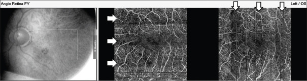
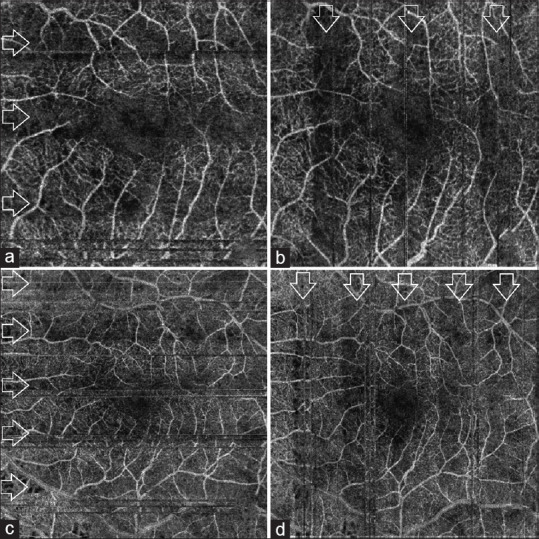
Methods: Seven primary open-angle glaucoma patients (eight eyes) were included, with a median age of 67.0 years (range, 39-73 years), who demonstrated alternating hypointense bands of OCTA flow signal on the macular scan at increased intraocular pressure (IOP). All patients received comprehensive ophthalmic examination, OCTA examination with RTVue-XR, and infrared video scanning laser ophthalmoscopy. Changes in retinal microcirculation were assessed on the raw OCTA scans as well as the resultant vessel density maps before and after IOP reduction.
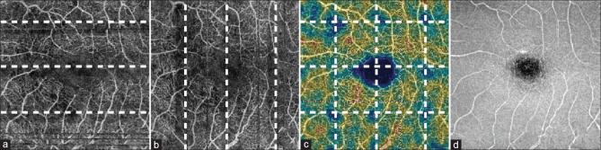
Results: Median IOP in study eyes was 39.0 mmHg (range, 36-58 mmHg). Hypointense bands of OCTA flow signal were associated with arterial pulsation on video scanning laser ophthalmoscopy in all eyes and agreed with the heart rate and resulted in a spotted grid pattern of hypoperfusion on vessel density maps in seven eyes. Median vessel density in superficial capillary plexus and deep capillary plexus was 32.4% and 47.2%, respectively, at high IOP, and increased statistically significantly to 36.5% (P = 0.016) and 50.9% (P = 0.016), respectively, after IOP reduction.
Conclusions: Alternating hypointense flow signal bands on OCTA scans are possibly caused by the pulsatile character of retinal blood flow during the cardiac cycle in eyes with high IOP and may reflect the imbalance between IOP and perfusion pressure. This phenomenon is responsible for the reversible decrease of vessel density at high IOP.
期刊介绍:
Peer Review under the responsibility of Iranian Society of Ophthalmology Journal of Current Ophthalmology, the official publication of the Iranian Society of Ophthalmology, is a peer-reviewed, open-access, scientific journal that welcomes high quality original articles related to vision science and all fields of ophthalmology. Journal of Current Ophthalmology is the continuum of Iranian Journal of Ophthalmology published since 1969.

 求助内容:
求助内容: 应助结果提醒方式:


