{"title":"腕管综合征患者手腕MRI和正中神经扩散张量成像。","authors":"Suprava Naik, Siladitya Mahanty, Sanjeev Kumar Bhoi, Yuvraj Lahre, Nerbadyswari Deep Bag, Sudipta Mohakud","doi":"10.25259/JNRP_57_2022","DOIUrl":null,"url":null,"abstract":"<p><strong>Objectives: </strong>Diagnosis of carpal tunnel syndrome (CTS) is based on the clinical symptoms and nerve conduction study. Magnetic resonance imaging (MRI) is non-invasive objective tool for assessing the median nerve and carpal tunnel. The purpose of this study was to evaluate MRI changes in patients with CTS, and compare them with healthy subjects.</p><p><strong>Materials and methods: </strong>Forty-three CTS patients and 43 age matched control were included and scanned in a 3T MRI scanner. Cross-sectional areas (CSA) of median nerve were measured at the level of distal radio-ulnar joint level (CSA1), proximal row of carpal bone (CSA2), and hook of hamate (CSA3). Flattening ratio (FR) of median nerve, thickness of flexor retinaculum, median nerve signal intensity, and thenar muscles were assessed. Fractional anisotropy (FA), average diffusion coefficient (ADC), and radial diffusivity (RD) of median nerve of CTS patients were obtained from diffusion tensor imaging (DTI) and compared with those of controls.</p><p><strong>Results: </strong>Thirty-three patients (76.7%) were female. Mean duration of the pain was 7.4 ± 2.6 months. The mean CSA1 (13.2 ± 4.2 mm<sup>2</sup>), CSA2 (12.5 ± 3.5 mm<sup>2</sup>), and CSA3 (9.2 ± 1.5 mm<sup>2</sup>) in CTS patients were significantly higher compared to control group: CSA1 (10.15 ± 1.64 mm<sup>2</sup>), CSA2 (9.38 ± 1.37 mm<sup>2</sup>), and CSA3 (8.4 ± 0.9 mm<sup>2</sup>), (<i>P</i> = 0.001 in all). The mean FR of median nerve and thickness of flexor retinaculum were increased in CTS patients. The mean FA was reduced in CTS patients compared to control proximal to carpal tunnel and within the tunnel. Mean ADC and RD values were higher in CTS patients as compared to control for both levels.</p><p><strong>Conclusion: </strong>MRI can detect subtle changes in the median nerve and thenar muscles in CTS and may be useful in equivocal cases and to exclude secondary causes of CTS. DTI shows reduced FA and increased ADC and RD in CTS patients.</p>","PeriodicalId":16443,"journal":{"name":"Journal of Neurosciences in Rural Practice","volume":"14 2","pages":"302-307"},"PeriodicalIF":0.9000,"publicationDate":"2023-04-01","publicationTypes":"Journal Article","fieldsOfStudy":null,"isOpenAccess":false,"openAccessPdf":"https://www.ncbi.nlm.nih.gov/pmc/articles/PMC10174157/pdf/","citationCount":"0","resultStr":"{\"title\":\"MRI of wrist and diffusion tensor imaging of the median nerve in patients with carpal tunnel syndrome.\",\"authors\":\"Suprava Naik, Siladitya Mahanty, Sanjeev Kumar Bhoi, Yuvraj Lahre, Nerbadyswari Deep Bag, Sudipta Mohakud\",\"doi\":\"10.25259/JNRP_57_2022\",\"DOIUrl\":null,\"url\":null,\"abstract\":\"<p><strong>Objectives: </strong>Diagnosis of carpal tunnel syndrome (CTS) is based on the clinical symptoms and nerve conduction study. Magnetic resonance imaging (MRI) is non-invasive objective tool for assessing the median nerve and carpal tunnel. The purpose of this study was to evaluate MRI changes in patients with CTS, and compare them with healthy subjects.</p><p><strong>Materials and methods: </strong>Forty-three CTS patients and 43 age matched control were included and scanned in a 3T MRI scanner. Cross-sectional areas (CSA) of median nerve were measured at the level of distal radio-ulnar joint level (CSA1), proximal row of carpal bone (CSA2), and hook of hamate (CSA3). Flattening ratio (FR) of median nerve, thickness of flexor retinaculum, median nerve signal intensity, and thenar muscles were assessed. Fractional anisotropy (FA), average diffusion coefficient (ADC), and radial diffusivity (RD) of median nerve of CTS patients were obtained from diffusion tensor imaging (DTI) and compared with those of controls.</p><p><strong>Results: </strong>Thirty-three patients (76.7%) were female. Mean duration of the pain was 7.4 ± 2.6 months. The mean CSA1 (13.2 ± 4.2 mm<sup>2</sup>), CSA2 (12.5 ± 3.5 mm<sup>2</sup>), and CSA3 (9.2 ± 1.5 mm<sup>2</sup>) in CTS patients were significantly higher compared to control group: CSA1 (10.15 ± 1.64 mm<sup>2</sup>), CSA2 (9.38 ± 1.37 mm<sup>2</sup>), and CSA3 (8.4 ± 0.9 mm<sup>2</sup>), (<i>P</i> = 0.001 in all). The mean FR of median nerve and thickness of flexor retinaculum were increased in CTS patients. The mean FA was reduced in CTS patients compared to control proximal to carpal tunnel and within the tunnel. Mean ADC and RD values were higher in CTS patients as compared to control for both levels.</p><p><strong>Conclusion: </strong>MRI can detect subtle changes in the median nerve and thenar muscles in CTS and may be useful in equivocal cases and to exclude secondary causes of CTS. DTI shows reduced FA and increased ADC and RD in CTS patients.</p>\",\"PeriodicalId\":16443,\"journal\":{\"name\":\"Journal of Neurosciences in Rural Practice\",\"volume\":\"14 2\",\"pages\":\"302-307\"},\"PeriodicalIF\":0.9000,\"publicationDate\":\"2023-04-01\",\"publicationTypes\":\"Journal Article\",\"fieldsOfStudy\":null,\"isOpenAccess\":false,\"openAccessPdf\":\"https://www.ncbi.nlm.nih.gov/pmc/articles/PMC10174157/pdf/\",\"citationCount\":\"0\",\"resultStr\":null,\"platform\":\"Semanticscholar\",\"paperid\":null,\"PeriodicalName\":\"Journal of Neurosciences in Rural Practice\",\"FirstCategoryId\":\"1085\",\"ListUrlMain\":\"https://doi.org/10.25259/JNRP_57_2022\",\"RegionNum\":0,\"RegionCategory\":null,\"ArticlePicture\":[],\"TitleCN\":null,\"AbstractTextCN\":null,\"PMCID\":null,\"EPubDate\":\"2023/3/16 0:00:00\",\"PubModel\":\"Epub\",\"JCR\":\"Q4\",\"JCRName\":\"CLINICAL NEUROLOGY\",\"Score\":null,\"Total\":0}","platform":"Semanticscholar","paperid":null,"PeriodicalName":"Journal of Neurosciences in Rural Practice","FirstCategoryId":"1085","ListUrlMain":"https://doi.org/10.25259/JNRP_57_2022","RegionNum":0,"RegionCategory":null,"ArticlePicture":[],"TitleCN":null,"AbstractTextCN":null,"PMCID":null,"EPubDate":"2023/3/16 0:00:00","PubModel":"Epub","JCR":"Q4","JCRName":"CLINICAL NEUROLOGY","Score":null,"Total":0}
MRI of wrist and diffusion tensor imaging of the median nerve in patients with carpal tunnel syndrome.
Objectives: Diagnosis of carpal tunnel syndrome (CTS) is based on the clinical symptoms and nerve conduction study. Magnetic resonance imaging (MRI) is non-invasive objective tool for assessing the median nerve and carpal tunnel. The purpose of this study was to evaluate MRI changes in patients with CTS, and compare them with healthy subjects.
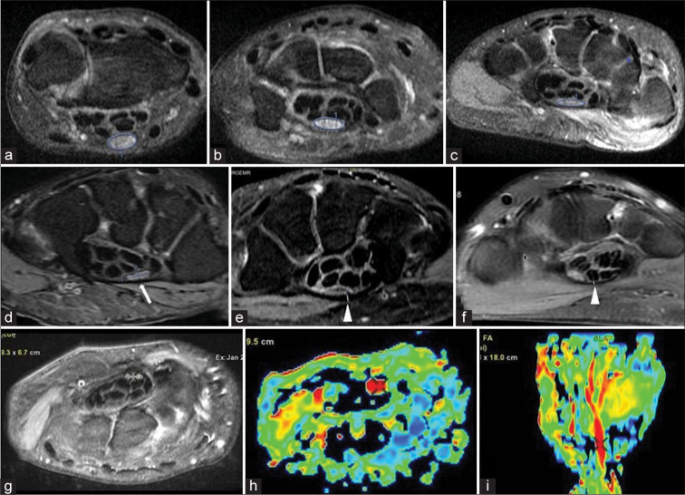
Materials and methods: Forty-three CTS patients and 43 age matched control were included and scanned in a 3T MRI scanner. Cross-sectional areas (CSA) of median nerve were measured at the level of distal radio-ulnar joint level (CSA1), proximal row of carpal bone (CSA2), and hook of hamate (CSA3). Flattening ratio (FR) of median nerve, thickness of flexor retinaculum, median nerve signal intensity, and thenar muscles were assessed. Fractional anisotropy (FA), average diffusion coefficient (ADC), and radial diffusivity (RD) of median nerve of CTS patients were obtained from diffusion tensor imaging (DTI) and compared with those of controls.
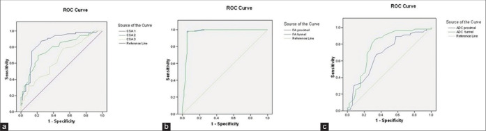
Results: Thirty-three patients (76.7%) were female. Mean duration of the pain was 7.4 ± 2.6 months. The mean CSA1 (13.2 ± 4.2 mm2), CSA2 (12.5 ± 3.5 mm2), and CSA3 (9.2 ± 1.5 mm2) in CTS patients were significantly higher compared to control group: CSA1 (10.15 ± 1.64 mm2), CSA2 (9.38 ± 1.37 mm2), and CSA3 (8.4 ± 0.9 mm2), (P = 0.001 in all). The mean FR of median nerve and thickness of flexor retinaculum were increased in CTS patients. The mean FA was reduced in CTS patients compared to control proximal to carpal tunnel and within the tunnel. Mean ADC and RD values were higher in CTS patients as compared to control for both levels.
Conclusion: MRI can detect subtle changes in the median nerve and thenar muscles in CTS and may be useful in equivocal cases and to exclude secondary causes of CTS. DTI shows reduced FA and increased ADC and RD in CTS patients.

 求助内容:
求助内容: 应助结果提醒方式:


