Joseph Kyu-Hyung Park, Se Yeon Lee, Jong-Ho Kim, Baek-Kyu Kim
{"title":"颧腋区纤维发育不良取芯术后的远期疗效。","authors":"Joseph Kyu-Hyung Park, Se Yeon Lee, Jong-Ho Kim, Baek-Kyu Kim","doi":"10.7181/acfs.2023.00038","DOIUrl":null,"url":null,"abstract":"<p><strong>Background: </strong>Fibrous dysplasia (FD) is a localized bone disorder in which fibro-osseous tissue replaces the normal bone structure. Patients with craniofacial FD often present with gradual swelling, deformity, and compromised vision or hearing. We previously introduced \"the core extirpation method,\" a novel surgical technique that is minimally invasive like traditional bone shaving but has longer-lasting effects. This study presents the long-term outcomes of our core extirpation method.</p><p><strong>Methods: </strong>We conducted a retrospective analysis of patients who underwent core extirpation for FD of the zygomaticomaxillary region from 2012 through 2021. Computed tomography (CT) scans were performed 6 to 12 months before the operation, immediately before and after the operation, and during follow-up visits. We performed all operations using the upper gingivobuccal approach, and we extirpated the core of the lesion while preserving the cortical structures of the zygoma and the maxilla to maintain symmetrical facial contour.</p><p><strong>Results: </strong>In 12 patients with lesions in the growth phase, anteroposterior/mediolateral (AP/ML) length discrepancies and the volume increased between preoperative and immediate postoperative CT scans. All patients' immediate postoperative AP/ML discrepancies were stable up to 12-17 months postoperatively. Postoperative volume showed continuous lesion growth; the median volume growth rate was 0.61 cc per month.</p><p><strong>Conclusion: </strong>In this article, we present our experiences managing FD using the minimally invasive core extirpation technique, which entails small expected blood loss and can be performed as day surgery. It provides similar cosmetic outcomes as traditional bone shaving but with longer-lasting results. Although there are some limitations with the study's retrospective nature and small sample size, our 4-year follow-up results show promising results of the core extirpation method in well-indicated patients.</p>","PeriodicalId":52238,"journal":{"name":"Archives of Craniofacial Surgery","volume":"24 2","pages":"59-65"},"PeriodicalIF":0.0000,"publicationDate":"2023-04-01","publicationTypes":"Journal Article","fieldsOfStudy":null,"isOpenAccess":false,"openAccessPdf":"https://ftp.ncbi.nlm.nih.gov/pub/pmc/oa_pdf/b0/03/acfs-2023-00038.PMC10165236.pdf","citationCount":"1","resultStr":"{\"title\":\"Long-term outcomes after core extirpation of fibrous dysplasia of the zygomaticomaxillary region.\",\"authors\":\"Joseph Kyu-Hyung Park, Se Yeon Lee, Jong-Ho Kim, Baek-Kyu Kim\",\"doi\":\"10.7181/acfs.2023.00038\",\"DOIUrl\":null,\"url\":null,\"abstract\":\"<p><strong>Background: </strong>Fibrous dysplasia (FD) is a localized bone disorder in which fibro-osseous tissue replaces the normal bone structure. Patients with craniofacial FD often present with gradual swelling, deformity, and compromised vision or hearing. We previously introduced \\\"the core extirpation method,\\\" a novel surgical technique that is minimally invasive like traditional bone shaving but has longer-lasting effects. This study presents the long-term outcomes of our core extirpation method.</p><p><strong>Methods: </strong>We conducted a retrospective analysis of patients who underwent core extirpation for FD of the zygomaticomaxillary region from 2012 through 2021. Computed tomography (CT) scans were performed 6 to 12 months before the operation, immediately before and after the operation, and during follow-up visits. We performed all operations using the upper gingivobuccal approach, and we extirpated the core of the lesion while preserving the cortical structures of the zygoma and the maxilla to maintain symmetrical facial contour.</p><p><strong>Results: </strong>In 12 patients with lesions in the growth phase, anteroposterior/mediolateral (AP/ML) length discrepancies and the volume increased between preoperative and immediate postoperative CT scans. All patients' immediate postoperative AP/ML discrepancies were stable up to 12-17 months postoperatively. Postoperative volume showed continuous lesion growth; the median volume growth rate was 0.61 cc per month.</p><p><strong>Conclusion: </strong>In this article, we present our experiences managing FD using the minimally invasive core extirpation technique, which entails small expected blood loss and can be performed as day surgery. It provides similar cosmetic outcomes as traditional bone shaving but with longer-lasting results. Although there are some limitations with the study's retrospective nature and small sample size, our 4-year follow-up results show promising results of the core extirpation method in well-indicated patients.</p>\",\"PeriodicalId\":52238,\"journal\":{\"name\":\"Archives of Craniofacial Surgery\",\"volume\":\"24 2\",\"pages\":\"59-65\"},\"PeriodicalIF\":0.0000,\"publicationDate\":\"2023-04-01\",\"publicationTypes\":\"Journal Article\",\"fieldsOfStudy\":null,\"isOpenAccess\":false,\"openAccessPdf\":\"https://ftp.ncbi.nlm.nih.gov/pub/pmc/oa_pdf/b0/03/acfs-2023-00038.PMC10165236.pdf\",\"citationCount\":\"1\",\"resultStr\":null,\"platform\":\"Semanticscholar\",\"paperid\":null,\"PeriodicalName\":\"Archives of Craniofacial Surgery\",\"FirstCategoryId\":\"1085\",\"ListUrlMain\":\"https://doi.org/10.7181/acfs.2023.00038\",\"RegionNum\":0,\"RegionCategory\":null,\"ArticlePicture\":[],\"TitleCN\":null,\"AbstractTextCN\":null,\"PMCID\":null,\"EPubDate\":\"\",\"PubModel\":\"\",\"JCR\":\"Q2\",\"JCRName\":\"Medicine\",\"Score\":null,\"Total\":0}","platform":"Semanticscholar","paperid":null,"PeriodicalName":"Archives of Craniofacial Surgery","FirstCategoryId":"1085","ListUrlMain":"https://doi.org/10.7181/acfs.2023.00038","RegionNum":0,"RegionCategory":null,"ArticlePicture":[],"TitleCN":null,"AbstractTextCN":null,"PMCID":null,"EPubDate":"","PubModel":"","JCR":"Q2","JCRName":"Medicine","Score":null,"Total":0}
Long-term outcomes after core extirpation of fibrous dysplasia of the zygomaticomaxillary region.
Background: Fibrous dysplasia (FD) is a localized bone disorder in which fibro-osseous tissue replaces the normal bone structure. Patients with craniofacial FD often present with gradual swelling, deformity, and compromised vision or hearing. We previously introduced "the core extirpation method," a novel surgical technique that is minimally invasive like traditional bone shaving but has longer-lasting effects. This study presents the long-term outcomes of our core extirpation method.
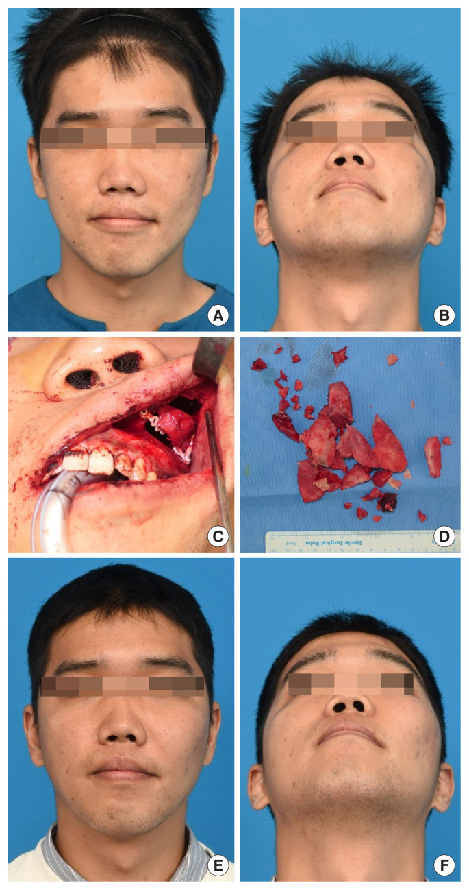
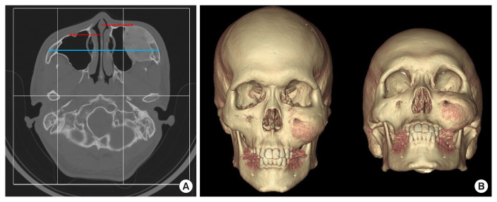
Methods: We conducted a retrospective analysis of patients who underwent core extirpation for FD of the zygomaticomaxillary region from 2012 through 2021. Computed tomography (CT) scans were performed 6 to 12 months before the operation, immediately before and after the operation, and during follow-up visits. We performed all operations using the upper gingivobuccal approach, and we extirpated the core of the lesion while preserving the cortical structures of the zygoma and the maxilla to maintain symmetrical facial contour.
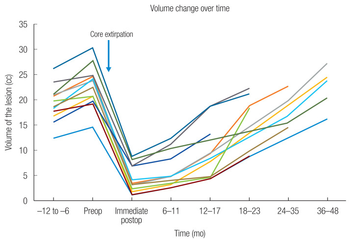
Results: In 12 patients with lesions in the growth phase, anteroposterior/mediolateral (AP/ML) length discrepancies and the volume increased between preoperative and immediate postoperative CT scans. All patients' immediate postoperative AP/ML discrepancies were stable up to 12-17 months postoperatively. Postoperative volume showed continuous lesion growth; the median volume growth rate was 0.61 cc per month.
Conclusion: In this article, we present our experiences managing FD using the minimally invasive core extirpation technique, which entails small expected blood loss and can be performed as day surgery. It provides similar cosmetic outcomes as traditional bone shaving but with longer-lasting results. Although there are some limitations with the study's retrospective nature and small sample size, our 4-year follow-up results show promising results of the core extirpation method in well-indicated patients.

 求助内容:
求助内容: 应助结果提醒方式:


