{"title":"黏膜内黑素细胞痣-牙龈肿大的罕见原因。个案报告及文献回顾。","authors":"Roshni Ramesh, Arun Sadasivan","doi":"10.2147/CCIDE.S408425","DOIUrl":null,"url":null,"abstract":"<p><strong>Background: </strong>Oral melanocytic nevi are infrequent oral lesions derived from nevus cells of oral mucosa which causes focal hyperpigmentation. The most common site of occurrence of oral nevi is the hard palate followed by buccal mucosa and gingiva. The mean age group affected are in their 3rd and 4th decade of life and there seems to be a predilection for females. Clinically, oral nevi are usually small, well-circumscribed macules but can also present as slightly raised papules. Histologically, nevi can be classified as Junctional, Compound or Intramucosal, with intramucosal being the more common type in the oral cavity.</p><p><strong>Case presentation: </strong>In this paper, we report a case of intramucosal nevus in a 25-year-old female patient. The lesion presented as a gingival enlargement in the mandibular anterior region involving the marginal and attached gingiva, which is an extremely rare presentation. The clinical findings, histologic features and surgical management are presented. The patient was followed up for one year and the one year follow up revealed a small area of focal hyperpigmentation at the site of the previous lesion which is being closely monitored.</p><p><strong>Conclusion: </strong>Nevi located in the mucous membrane have been documented to pose a threat of malignant transformation. Hence, all pigmented lesions of the oral cavity should be cautiously diagnosed.</p>","PeriodicalId":10445,"journal":{"name":"Clinical, Cosmetic and Investigational Dentistry","volume":"15 ","pages":"71-77"},"PeriodicalIF":1.5000,"publicationDate":"2023-01-01","publicationTypes":"Journal Article","fieldsOfStudy":null,"isOpenAccess":false,"openAccessPdf":"https://ftp.ncbi.nlm.nih.gov/pub/pmc/oa_pdf/38/c9/ccide-15-71.PMC10155718.pdf","citationCount":"0","resultStr":"{\"title\":\"Intramucosal Melanocytic Nevi - A Rare Cause for Gingival Enlargement. Report of a Case and Review of Literature.\",\"authors\":\"Roshni Ramesh, Arun Sadasivan\",\"doi\":\"10.2147/CCIDE.S408425\",\"DOIUrl\":null,\"url\":null,\"abstract\":\"<p><strong>Background: </strong>Oral melanocytic nevi are infrequent oral lesions derived from nevus cells of oral mucosa which causes focal hyperpigmentation. The most common site of occurrence of oral nevi is the hard palate followed by buccal mucosa and gingiva. The mean age group affected are in their 3rd and 4th decade of life and there seems to be a predilection for females. Clinically, oral nevi are usually small, well-circumscribed macules but can also present as slightly raised papules. Histologically, nevi can be classified as Junctional, Compound or Intramucosal, with intramucosal being the more common type in the oral cavity.</p><p><strong>Case presentation: </strong>In this paper, we report a case of intramucosal nevus in a 25-year-old female patient. The lesion presented as a gingival enlargement in the mandibular anterior region involving the marginal and attached gingiva, which is an extremely rare presentation. The clinical findings, histologic features and surgical management are presented. The patient was followed up for one year and the one year follow up revealed a small area of focal hyperpigmentation at the site of the previous lesion which is being closely monitored.</p><p><strong>Conclusion: </strong>Nevi located in the mucous membrane have been documented to pose a threat of malignant transformation. Hence, all pigmented lesions of the oral cavity should be cautiously diagnosed.</p>\",\"PeriodicalId\":10445,\"journal\":{\"name\":\"Clinical, Cosmetic and Investigational Dentistry\",\"volume\":\"15 \",\"pages\":\"71-77\"},\"PeriodicalIF\":1.5000,\"publicationDate\":\"2023-01-01\",\"publicationTypes\":\"Journal Article\",\"fieldsOfStudy\":null,\"isOpenAccess\":false,\"openAccessPdf\":\"https://ftp.ncbi.nlm.nih.gov/pub/pmc/oa_pdf/38/c9/ccide-15-71.PMC10155718.pdf\",\"citationCount\":\"0\",\"resultStr\":null,\"platform\":\"Semanticscholar\",\"paperid\":null,\"PeriodicalName\":\"Clinical, Cosmetic and Investigational Dentistry\",\"FirstCategoryId\":\"1085\",\"ListUrlMain\":\"https://doi.org/10.2147/CCIDE.S408425\",\"RegionNum\":0,\"RegionCategory\":null,\"ArticlePicture\":[],\"TitleCN\":null,\"AbstractTextCN\":null,\"PMCID\":null,\"EPubDate\":\"\",\"PubModel\":\"\",\"JCR\":\"Q3\",\"JCRName\":\"DENTISTRY, ORAL SURGERY & MEDICINE\",\"Score\":null,\"Total\":0}","platform":"Semanticscholar","paperid":null,"PeriodicalName":"Clinical, Cosmetic and Investigational Dentistry","FirstCategoryId":"1085","ListUrlMain":"https://doi.org/10.2147/CCIDE.S408425","RegionNum":0,"RegionCategory":null,"ArticlePicture":[],"TitleCN":null,"AbstractTextCN":null,"PMCID":null,"EPubDate":"","PubModel":"","JCR":"Q3","JCRName":"DENTISTRY, ORAL SURGERY & MEDICINE","Score":null,"Total":0}
Intramucosal Melanocytic Nevi - A Rare Cause for Gingival Enlargement. Report of a Case and Review of Literature.
Background: Oral melanocytic nevi are infrequent oral lesions derived from nevus cells of oral mucosa which causes focal hyperpigmentation. The most common site of occurrence of oral nevi is the hard palate followed by buccal mucosa and gingiva. The mean age group affected are in their 3rd and 4th decade of life and there seems to be a predilection for females. Clinically, oral nevi are usually small, well-circumscribed macules but can also present as slightly raised papules. Histologically, nevi can be classified as Junctional, Compound or Intramucosal, with intramucosal being the more common type in the oral cavity.
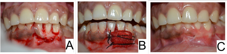
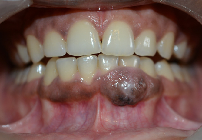
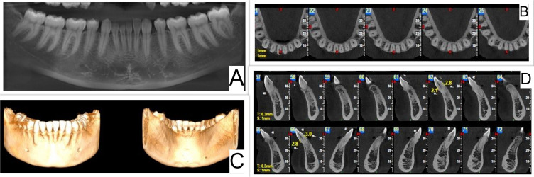
Case presentation: In this paper, we report a case of intramucosal nevus in a 25-year-old female patient. The lesion presented as a gingival enlargement in the mandibular anterior region involving the marginal and attached gingiva, which is an extremely rare presentation. The clinical findings, histologic features and surgical management are presented. The patient was followed up for one year and the one year follow up revealed a small area of focal hyperpigmentation at the site of the previous lesion which is being closely monitored.
Conclusion: Nevi located in the mucous membrane have been documented to pose a threat of malignant transformation. Hence, all pigmented lesions of the oral cavity should be cautiously diagnosed.

 求助内容:
求助内容: 应助结果提醒方式:


