Bhavna Jha Kukreja, Kishore G Bhat, Pankaj Kukreja, Ajaykumar Nayak, Vijaylakshmi S Kotrashetty, Santosh Dindawar, Rajkumar Balakrishnan
{"title":"动物模型中微型牙种植体周围牙周膜纤维的再生及其与骨骼的连接:一项放射学和组织学研究。","authors":"Bhavna Jha Kukreja, Kishore G Bhat, Pankaj Kukreja, Ajaykumar Nayak, Vijaylakshmi S Kotrashetty, Santosh Dindawar, Rajkumar Balakrishnan","doi":"10.4103/jisp.jisp_314_22","DOIUrl":null,"url":null,"abstract":"<p><strong>Background: </strong>Tissue-engineered periodontal ligament (PDL) around a dental implant by using PDL stem cells (PDLSCs) may be useful in periodontal regeneration and can reduce or eliminate certain shortcomings of dental implants.</p><p><strong>Materials and methods: </strong>PDLSCs were isolated from extracted human PDL cells and cultured in a bioreactor. They were identified using markers CD45, CD73, CD90, CD105, and CD146. After the formation of multiple cellular layers, they were then attached on titanium mini dental implants and placed in rabbit tibia. The rabbits were sacrificed after 9 months, and the implants were analyzed histologically and radiographically by Cone beam computed tomography (CBCT).</p><p><strong>Results: </strong>Isolated PDLSCs obtained from human premolars showed a colony-forming ability on the 7<sup>th</sup> day and 14<sup>th</sup> day. Immunocytochemistry revealed that cells had taken up the adequate positive stains for primary antibodies CD73, CD90, CD105, and CD146 and negative staining for CD45. The histological sections obtained from sacrificed rabbits, when viewed under the light microscope, clearly showed the presence of PDL around dental implants. CBCT examination showed that the implant was well within the bone and did not migrate. The site appeared to be normal without any lytic changes in the bone.</p><p><strong>Conclusion: </strong>It can safely be postulated from the present study that tissue engineering of PDL can be achieved around dental implants using PDLSCs. Important inter-tissue interactions like the formation of a functional PDL around the implantation site, and induction of bone formation in the vicinity of the implants may be possible. Future research in humans is required for further research.</p>","PeriodicalId":15890,"journal":{"name":"Journal of Indian Society of Periodontology","volume":null,"pages":null},"PeriodicalIF":0.0000,"publicationDate":"2023-03-01","publicationTypes":"Journal Article","fieldsOfStudy":null,"isOpenAccess":false,"openAccessPdf":"https://www.ncbi.nlm.nih.gov/pmc/articles/PMC10159091/pdf/","citationCount":"0","resultStr":"{\"title\":\"Regeneration of periodontal ligament fibers around mini dental implants and their attachment to the bone in an animal model: A radiographic and histological study.\",\"authors\":\"Bhavna Jha Kukreja, Kishore G Bhat, Pankaj Kukreja, Ajaykumar Nayak, Vijaylakshmi S Kotrashetty, Santosh Dindawar, Rajkumar Balakrishnan\",\"doi\":\"10.4103/jisp.jisp_314_22\",\"DOIUrl\":null,\"url\":null,\"abstract\":\"<p><strong>Background: </strong>Tissue-engineered periodontal ligament (PDL) around a dental implant by using PDL stem cells (PDLSCs) may be useful in periodontal regeneration and can reduce or eliminate certain shortcomings of dental implants.</p><p><strong>Materials and methods: </strong>PDLSCs were isolated from extracted human PDL cells and cultured in a bioreactor. They were identified using markers CD45, CD73, CD90, CD105, and CD146. After the formation of multiple cellular layers, they were then attached on titanium mini dental implants and placed in rabbit tibia. The rabbits were sacrificed after 9 months, and the implants were analyzed histologically and radiographically by Cone beam computed tomography (CBCT).</p><p><strong>Results: </strong>Isolated PDLSCs obtained from human premolars showed a colony-forming ability on the 7<sup>th</sup> day and 14<sup>th</sup> day. Immunocytochemistry revealed that cells had taken up the adequate positive stains for primary antibodies CD73, CD90, CD105, and CD146 and negative staining for CD45. The histological sections obtained from sacrificed rabbits, when viewed under the light microscope, clearly showed the presence of PDL around dental implants. CBCT examination showed that the implant was well within the bone and did not migrate. The site appeared to be normal without any lytic changes in the bone.</p><p><strong>Conclusion: </strong>It can safely be postulated from the present study that tissue engineering of PDL can be achieved around dental implants using PDLSCs. Important inter-tissue interactions like the formation of a functional PDL around the implantation site, and induction of bone formation in the vicinity of the implants may be possible. Future research in humans is required for further research.</p>\",\"PeriodicalId\":15890,\"journal\":{\"name\":\"Journal of Indian Society of Periodontology\",\"volume\":null,\"pages\":null},\"PeriodicalIF\":0.0000,\"publicationDate\":\"2023-03-01\",\"publicationTypes\":\"Journal Article\",\"fieldsOfStudy\":null,\"isOpenAccess\":false,\"openAccessPdf\":\"https://www.ncbi.nlm.nih.gov/pmc/articles/PMC10159091/pdf/\",\"citationCount\":\"0\",\"resultStr\":null,\"platform\":\"Semanticscholar\",\"paperid\":null,\"PeriodicalName\":\"Journal of Indian Society of Periodontology\",\"FirstCategoryId\":\"1085\",\"ListUrlMain\":\"https://doi.org/10.4103/jisp.jisp_314_22\",\"RegionNum\":0,\"RegionCategory\":null,\"ArticlePicture\":[],\"TitleCN\":null,\"AbstractTextCN\":null,\"PMCID\":null,\"EPubDate\":\"2023/3/4 0:00:00\",\"PubModel\":\"Epub\",\"JCR\":\"Q2\",\"JCRName\":\"Dentistry\",\"Score\":null,\"Total\":0}","platform":"Semanticscholar","paperid":null,"PeriodicalName":"Journal of Indian Society of Periodontology","FirstCategoryId":"1085","ListUrlMain":"https://doi.org/10.4103/jisp.jisp_314_22","RegionNum":0,"RegionCategory":null,"ArticlePicture":[],"TitleCN":null,"AbstractTextCN":null,"PMCID":null,"EPubDate":"2023/3/4 0:00:00","PubModel":"Epub","JCR":"Q2","JCRName":"Dentistry","Score":null,"Total":0}
Regeneration of periodontal ligament fibers around mini dental implants and their attachment to the bone in an animal model: A radiographic and histological study.
Background: Tissue-engineered periodontal ligament (PDL) around a dental implant by using PDL stem cells (PDLSCs) may be useful in periodontal regeneration and can reduce or eliminate certain shortcomings of dental implants.
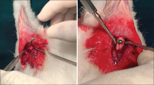
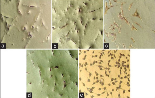
Materials and methods: PDLSCs were isolated from extracted human PDL cells and cultured in a bioreactor. They were identified using markers CD45, CD73, CD90, CD105, and CD146. After the formation of multiple cellular layers, they were then attached on titanium mini dental implants and placed in rabbit tibia. The rabbits were sacrificed after 9 months, and the implants were analyzed histologically and radiographically by Cone beam computed tomography (CBCT).
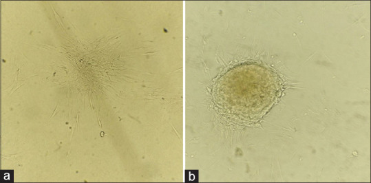
Results: Isolated PDLSCs obtained from human premolars showed a colony-forming ability on the 7th day and 14th day. Immunocytochemistry revealed that cells had taken up the adequate positive stains for primary antibodies CD73, CD90, CD105, and CD146 and negative staining for CD45. The histological sections obtained from sacrificed rabbits, when viewed under the light microscope, clearly showed the presence of PDL around dental implants. CBCT examination showed that the implant was well within the bone and did not migrate. The site appeared to be normal without any lytic changes in the bone.
Conclusion: It can safely be postulated from the present study that tissue engineering of PDL can be achieved around dental implants using PDLSCs. Important inter-tissue interactions like the formation of a functional PDL around the implantation site, and induction of bone formation in the vicinity of the implants may be possible. Future research in humans is required for further research.
期刊介绍:
The Journal of Indian Society of Periodontology publishes original scientific articles to support practice , education and research in the dental specialty of periodontology and oral implantology. Journal of Indian Society of Periodontology (JISP), is the official publication of the Society and is managed and brought out by the Editor of the society. The journal is published Bimonthly with special issues being brought out for specific occasions. The ISP had a bulletin as its publication for a large number of years and was enhanced as a Journal a few years ago

 求助内容:
求助内容: 应助结果提醒方式:


