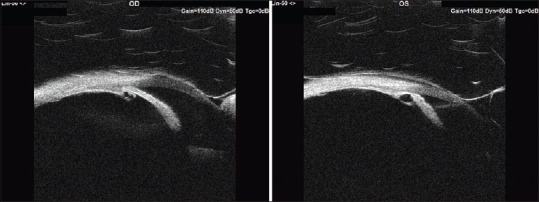
双侧多发睫状体囊肿合并闭角型青光眼1例。

IF 0.5 Q4 OPHTHALMOLOGY
Amar Almulhim, Ehab Alsirhy, Saleh A Al-Obeidan
{"title":"双侧多发睫状体囊肿合并闭角型青光眼1例。","authors":"Amar Almulhim,&nbsp;Ehab Alsirhy,&nbsp;Saleh A Al-Obeidan","doi":"10.4103/meajo.meajo_185_22","DOIUrl":null,"url":null,"abstract":"<p><p>We present the rare case of an 18-year-old medically free male who had a history of decrease in vision in the left eye (LE) in the last 4 years. On examination, best-corrected visual acuity was 20/20 in the right eye (RE) and counting fingers 3 feet in the LE. Intraocular pressure was 34 and 40 mmHg in RE and LE, respectively. Fundus examination showed cupping of 0.7 on the RE and 0.9 on the LE. Gonioscopy revealed bilateral angle closure with a double-hump sign. Ultrasound biomicroscopy showed multiple ciliary body cysts replacing ciliary body sulcus space bilaterally.</p>","PeriodicalId":18740,"journal":{"name":"Middle East African Journal of Ophthalmology","volume":"29 2","pages":"100-102"},"PeriodicalIF":0.5000,"publicationDate":"2022-04-01","publicationTypes":"Journal Article","fieldsOfStudy":null,"isOpenAccess":false,"openAccessPdf":"https://www.ncbi.nlm.nih.gov/pmc/articles/PMC10138131/pdf/","citationCount":"0","resultStr":"{\"title\":\"Bilateral Multiple Ciliary Body Cysts with Angle-Closure Glaucoma in an 18-Year-Old Patient.\",\"authors\":\"Amar Almulhim,&nbsp;Ehab Alsirhy,&nbsp;Saleh A Al-Obeidan\",\"doi\":\"10.4103/meajo.meajo_185_22\",\"DOIUrl\":null,\"url\":null,\"abstract\":\"<p><p>We present the rare case of an 18-year-old medically free male who had a history of decrease in vision in the left eye (LE) in the last 4 years. On examination, best-corrected visual acuity was 20/20 in the right eye (RE) and counting fingers 3 feet in the LE. Intraocular pressure was 34 and 40 mmHg in RE and LE, respectively. Fundus examination showed cupping of 0.7 on the RE and 0.9 on the LE. Gonioscopy revealed bilateral angle closure with a double-hump sign. Ultrasound biomicroscopy showed multiple ciliary body cysts replacing ciliary body sulcus space bilaterally.</p>\",\"PeriodicalId\":18740,\"journal\":{\"name\":\"Middle East African Journal of Ophthalmology\",\"volume\":\"29 2\",\"pages\":\"100-102\"},\"PeriodicalIF\":0.5000,\"publicationDate\":\"2022-04-01\",\"publicationTypes\":\"Journal Article\",\"fieldsOfStudy\":null,\"isOpenAccess\":false,\"openAccessPdf\":\"https://www.ncbi.nlm.nih.gov/pmc/articles/PMC10138131/pdf/\",\"citationCount\":\"0\",\"resultStr\":null,\"platform\":\"Semanticscholar\",\"paperid\":null,\"PeriodicalName\":\"Middle East African Journal of Ophthalmology\",\"FirstCategoryId\":\"1085\",\"ListUrlMain\":\"https://doi.org/10.4103/meajo.meajo_185_22\",\"RegionNum\":0,\"RegionCategory\":null,\"ArticlePicture\":[],\"TitleCN\":null,\"AbstractTextCN\":null,\"PMCID\":null,\"EPubDate\":\"\",\"PubModel\":\"\",\"JCR\":\"Q4\",\"JCRName\":\"OPHTHALMOLOGY\",\"Score\":null,\"Total\":0}","platform":"Semanticscholar","paperid":null,"PeriodicalName":"Middle East African Journal of Ophthalmology","FirstCategoryId":"1085","ListUrlMain":"https://doi.org/10.4103/meajo.meajo_185_22","RegionNum":0,"RegionCategory":null,"ArticlePicture":[],"TitleCN":null,"AbstractTextCN":null,"PMCID":null,"EPubDate":"","PubModel":"","JCR":"Q4","JCRName":"OPHTHALMOLOGY","Score":null,"Total":0}
引用次数: 0

摘要

我们提出一个罕见的情况下,18岁的男性医学自由谁有下降的历史,左眼视力(LE)在过去的4年。经检查,最佳矫正视力为右眼20/20,左眼数指3英尺。RE组和LE组眼压分别为34和40 mmHg。眼底检查显示RE为0.7,LE为0.9。角镜检查显示双侧角闭合伴双驼峰征。超声生物显微镜检查显示双侧多发睫状体囊肿取代睫状体沟间隙。
本文章由计算机程序翻译,如有差异,请以英文原文为准。

Bilateral Multiple Ciliary Body Cysts with Angle-Closure Glaucoma in an 18-Year-Old Patient.

Bilateral Multiple Ciliary Body Cysts with Angle-Closure Glaucoma in an 18-Year-Old Patient.

Bilateral Multiple Ciliary Body Cysts with Angle-Closure Glaucoma in an 18-Year-Old Patient.

Bilateral Multiple Ciliary Body Cysts with Angle-Closure Glaucoma in an 18-Year-Old Patient.

We present the rare case of an 18-year-old medically free male who had a history of decrease in vision in the left eye (LE) in the last 4 years. On examination, best-corrected visual acuity was 20/20 in the right eye (RE) and counting fingers 3 feet in the LE. Intraocular pressure was 34 and 40 mmHg in RE and LE, respectively. Fundus examination showed cupping of 0.7 on the RE and 0.9 on the LE. Gonioscopy revealed bilateral angle closure with a double-hump sign. Ultrasound biomicroscopy showed multiple ciliary body cysts replacing ciliary body sulcus space bilaterally.

求助全文
通过发布文献求助,成功后即可免费获取论文全文。 去求助
来源期刊
CiteScore
1.40
自引率
0.00%
发文量
1
期刊介绍: The Middle East African Journal of Ophthalmology (MEAJO), published four times per year in print and online, is an official journal of the Middle East African Council of Ophthalmology (MEACO). It is an international, peer-reviewed journal whose mission includes publication of original research of interest to ophthalmologists in the Middle East and Africa, and to provide readers with high quality educational review articles from world-renown experts. MEAJO, previously known as Middle East Journal of Ophthalmology (MEJO) was founded by Dr Akef El Maghraby in 1993.
×
引用
GB/T 7714-2015
复制
MLA
复制
APA
复制
导出至
BibTeX EndNote RefMan NoteFirst NoteExpress
×
提示
您的信息不完整,为了账户安全,请先补充。
现在去补充
×
提示
您因"违规操作"
具体请查看互助需知
我知道了
×
提示
确定
请完成安全验证×
copy
已复制链接
快去分享给好友吧!
我知道了
右上角分享
点击右上角分享
0
联系我们:info@booksci.cn Book学术提供免费学术资源搜索服务,方便国内外学者检索中英文文献。致力于提供最便捷和优质的服务体验。 Copyright © 2023 布克学术 All rights reserved.
京ICP备2023020795号-1
ghs 京公网安备 11010802042870号
Book学术文献互助
Book学术文献互助群
群 号:481959085
Book学术官方微信